

Why do super resolution and deep imaging seem to pull in opposite directions? In this fifth Quick Knowledge, we start from Abbe’s law and show how the numerical aperture (NA) and working distance (WD) of the objective set both the resolution and the imaging depth. We then compare the main super resolution (SR) techniques, from SMLM and STED to SIM and pixel reassignment, and rate how well each one suits live cells. Finally, we explain how Point REscan combines a more open pinhole with a highly sensitive camera to bring super resolution deep into living samples.
Quick Knowledge Part 5

DEEP LIVE CELL
IMAGING IN
SUPER
RESOLUTION

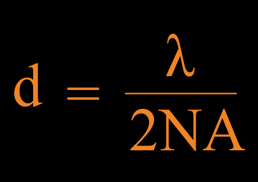
Equation: d = lambda divided by 2NA (Abbe diffraction limit)

Super
resolution

In theory, super resolution (SR) is
imaging beyond the diffraction limit
(d), defined by Abbe’s law as:

or

Ernst Abbe memorial
 Jena, Germany

Where d is the minimum resolvable distance, λ is the wavelength,
NA is the numerical aperture, n is the refractive index and α is the
maximum collection angle.

In practice, SR techniques are those that provide
resolution better than 200 nm...

...when imaged with an objective with high NA and
refractive index of oil. Usually, these objectives have
high magnification and short working distance (WD).

Sample

Super resolved image

Diffraction limited image

200 nm
SR dependent
on NA

According to Abbe’s law, d = λ/2NA,
the higher the NA, the better the
resolution.

Fibroblasts imaged in SR using Point REscan GAIA

30x
1.05NA

100x
1.50NA

NA defines the
angle of the
collected light, the
higher the NA, the
larger the angle and
the lower the
diffraction
limit (in nm)

Depth of SR
dependent on WD

Objectives with long working distance
(WD) have in general lower magnification
and lower NA than those with short WD.

Living zebrafish imaged in SR using Point REscan GAIA

the higher
the NA, the
shorter the
WD

30x
1.05NA

60x
1.30NA

0.8mmWD

0.3mmWD

Image depth 400 µm

Image depth 83 µm

Types of SR
techniques

Based on resolution, there are
two groups of SR techniques:

1. Providing resolution 100-200 nm:

2. Providing resolution beyond 100 nm:

PR (Pixel
reassignment)

d = 120-150 nm

SIM
d = 100-130 nm

FB (Fluctuation
based)

d = ca. 150 nm

ExM (Expansion
microscopy)

d = 50 nm

SMLM
d = 10-20 nm

STED
d = 50-100 nm

Jacquemet et al, J Cell Sci 2020
Zhang et al, Curr Protoc Neurosci 2020

SR techniques
not suitable for
living cells

The better the resolution of an SR
technique, the less suitable for live cell
imaging that technique is.

Live cell friendly

ExM

0/5, impossible due to the sample prep

SMLM

2/5, almost impossible due to the sample prep,
long acquisition and high laser powers

STED

3/5, possible only over very short periods due to
high laser powers

XY
resolution

Z
resolution

Thickness

Live cell
friendly

Image
fidelity

Multicolor

Temporal
resolution

Versatility

ExM
SMLM
STED

Jacquemet et al,
J Cell Sci 2020

SR techniques for
live cell imaging

Among all SR techniques, by far the best
ones for live cell imaging are those
based on pixel reassignment (PR).

Live cell friendly

PR

5/5, best suitable for live cell imaging

SIM

4/5, most common 3D SIM is more phototoxic than
TIRF SIM that is incapable for deep imaging

FB

4/5, phototoxicity varies depending on the
method (SOFI, SRRF)

XY
resolution

Z
resolution

Thickness

Live cell
friendly

Image
fidelity

Multicolor

Temporal
resolution

Versatility

PR
SIM
FB

Jacquemet et al,
J Cell Sci 2020

Deep live cell
imaging using PR
techniques

Point REscan stands out among pixel
reassignment techniques for its advantages
in both deep and live cell imaging.

1.

Being optimized for the widest range of objectives
gives REscan advantage in deep imaging as it
can use objectives with longer WD.

2.

More light through more open pinhole (≥1.5 AU)
and higher quantum efficiency of a detector (96%
QE) give REscan advantage in live cell imaging.

REscan collects
more light without
losing resolution.

Deep live cell
SR imaging

Solution to NA/WD problem in deep live cell SR
imaging is finding the optimal combination of
magnification, NA, pinhole size and camera.

GAIA is the solution that can provide live cell
imaging in SR deeper than 500 µm in sample.

GAIA pinhole size chart

M (magnification)

1.50
1.25
1.00
0.75
0.50
0.25
0.00

89
55
38
27
18

µm
µm
µm
µm
µm

-- 2 AU
- 1 AU

0

20

40

60

80

100

60x0.7

20x0.45

20x0.75

40x0.9

30x1.05

40x1.4

60x1.5

100x1.5

100x0.9

10x0.4

4x0.2

GAIA Point REscan super resolution confocal

Achieve the ideal ratio of objective NA & magnification, and
camera pixel size with our Line and Point REscan systems.
You can select the optimal pinhole size and image with a wide
range of objectives (4x-100x).

Download the quick knowledge

Receive more information about this knowledge article.