
NL5+

Fast 3D live cell imaging

Image thick samples with high contrast for long times.

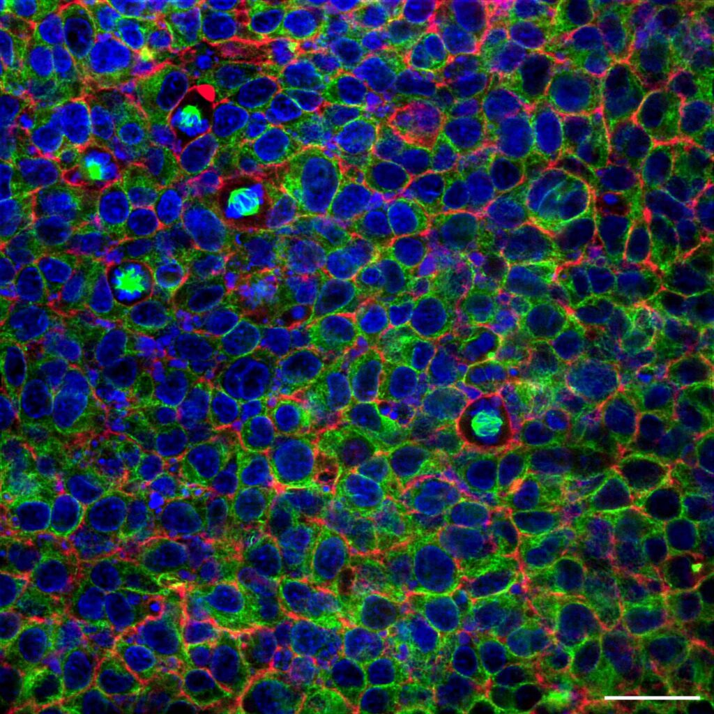
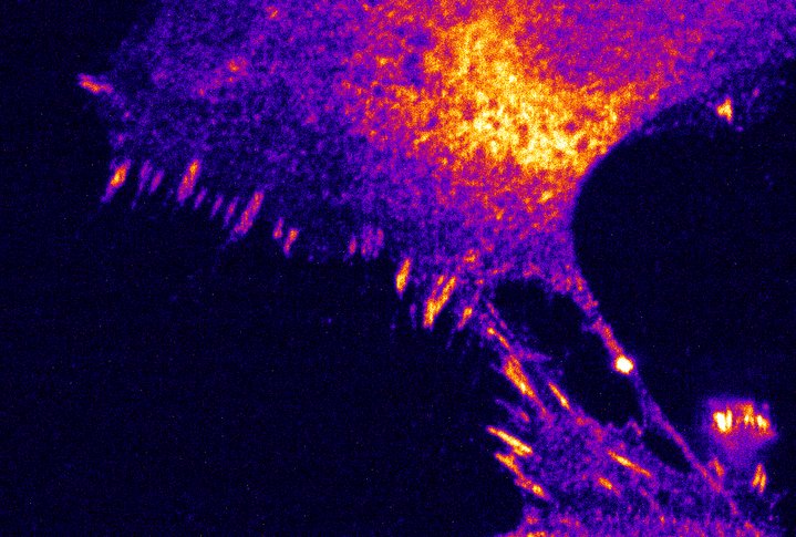
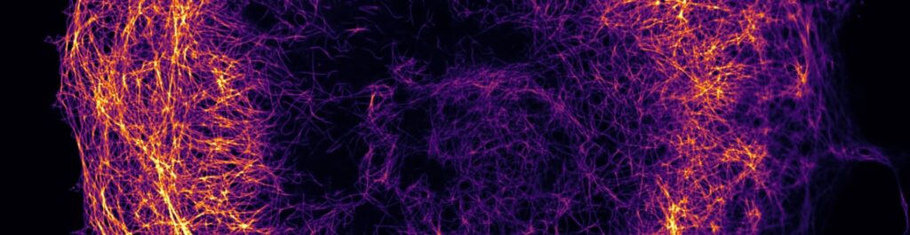
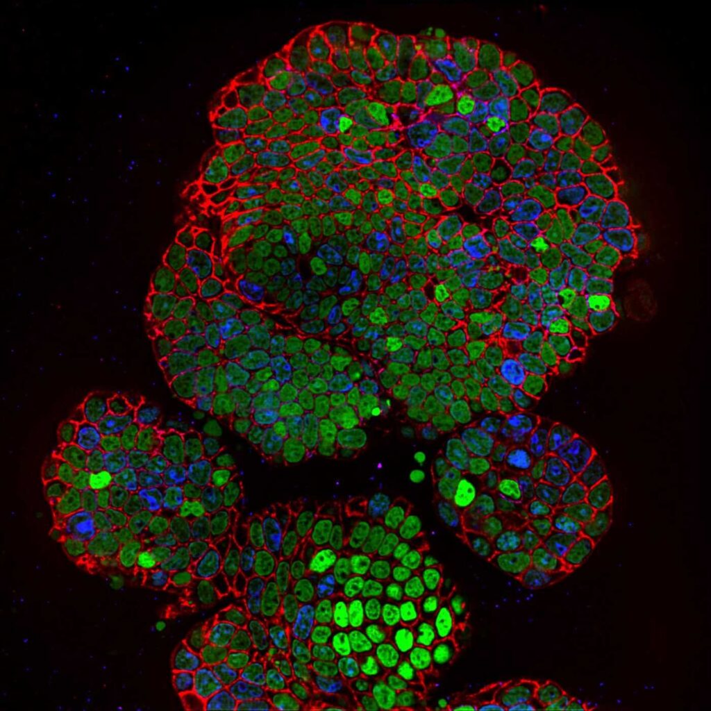
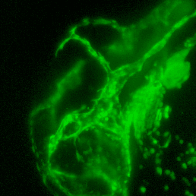
The NL5+ is the second generation of fast line-scanning confocal technologies. It provides high-contrast images from thicker specimens such as organoids, tissue samples, plant and animal model organisms. The gentle conditions for your live samples allow the NL5+ to excel in long time-lapse experiments and imaging low-signal samples.

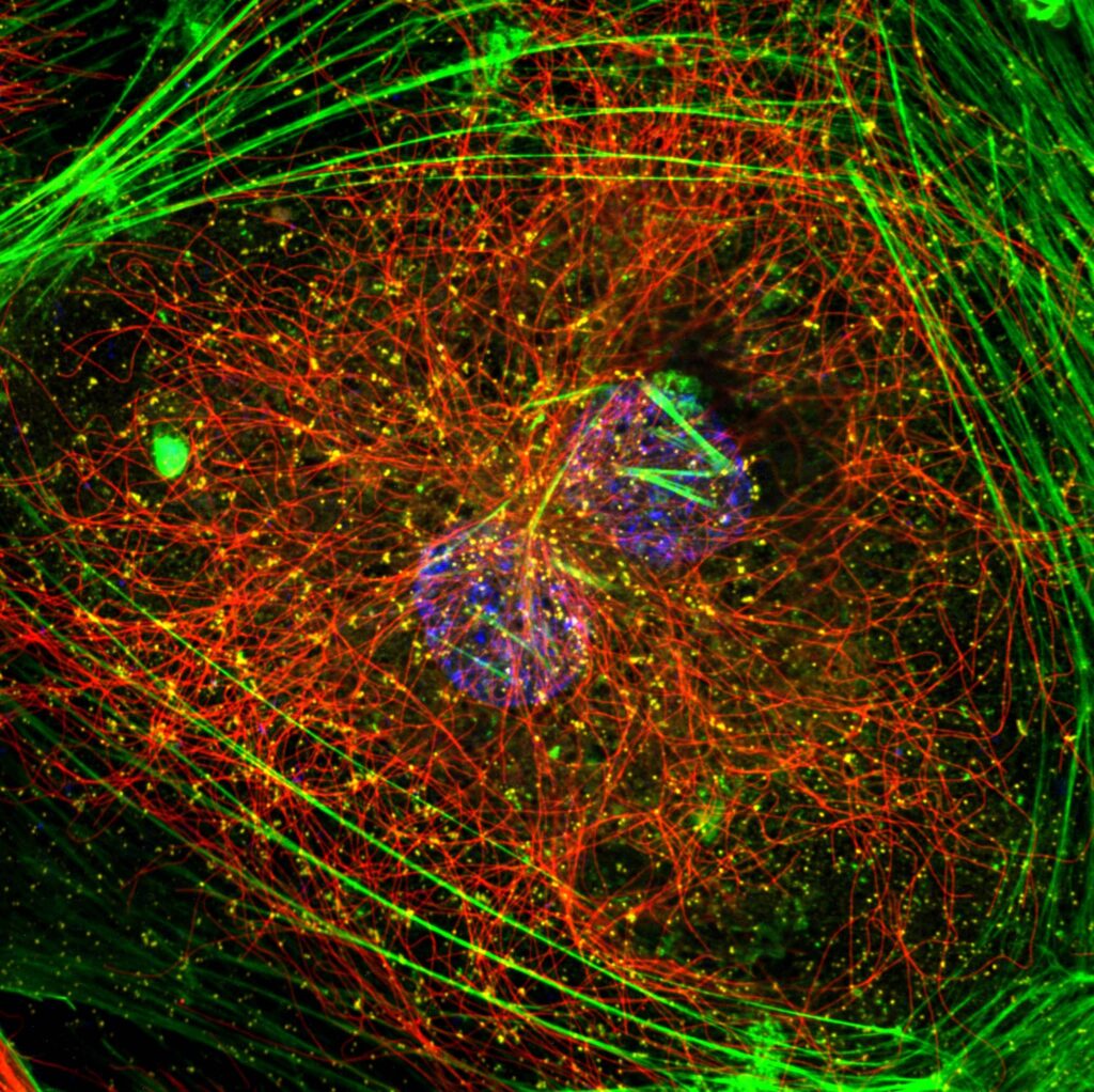
The combination of camera-based detection and a set of filters allows you to acquire your images in up to six different colors. This gives you an improved temporal resolution, high sensitivity and unprecedented signal-to-noise ratio. You get even faster imaging with cleaner results, thanks to additional motorized single-band emission filters.

3D live cell imaging with multiple filters

The NL5+ series takes the best of two worlds; point scanners and spinning disc, leading to a system with options to do confocal, widefield or brightfield observations.

Phototoxicity is a central problem in the application of confocal microscopy for live cell imaging. To overcome these challenges, we developed the second generation of fast line-scanning confocal technologies. Using a line scanner instead of a point scanner, a confocal microscope provides high temporal resolution, at the same time reducing the phototoxicity and allowing long-term imaging. A slit pinhole, together with an extremely sensitive sCMOS camera as a detector, makes the NL5+ series the perfect tool for fast and deep 3D live cell imaging.

Volvox autofluorescence 561 excitation channel. Image acquired 90 microns away from the coverslip 100x 1.5 objective was used with both systems

REscan

Fast line REscan confocal

Large FOV

Confocal imaging with large FOV

1 mm

Extra deep live cell imaging

90%

Less phototoxicity

No pinhole crosstalk

Contrary to spinning disk systems, another advantage of the NL5+ series and its slit pinhole is that it does not lead to pinhole crosstalk when imaging deep within the sample. The NL5+ series reaches 240 nm lateral resolution, which can be easily converted into 170 nm with on-the-fly deconvolution.

NL5+ is an excellent add-on to any research microscope. It allows converting any widefield system into a fast scanning confocal with high resolution and sensitivity.

The impact of an add-on filter

The NL5+ is a confocal system which enables even faster imaging with cleaner results. This is a direct result of one of the new functionalities: its additional motorized filter wheel.

Our system uses a line scanner, instead of a point scanner confocal microscope. This provides high temporal resolution, while at the same time reducing phototoxicity. Since phototoxicity is one of the biggest challenges for researchers, this makes the NL5+ the perfect tool for live cell imaging.

You can also add the NL5+ to any widefield microscope. This will turn it into an advanced fast-scanning confocal imaging system.

System

NL5+ technical specs

NL5+

$ 99

Per Month
Detector

Camera (sCMOS)


Resolution in real time

170 nm deconvolved,  240 nraw

Detector sensitivity
Up to 95% QE

FOV
FOV 18mm (FN18); 330×330 µm (40x objective)

Speed in line scanning
up to 75 fps for 2048×512 px and up to 50 fps for 2048×2048 px

Speed in fixed line mode
>1000 fps and more (camera limited)

Wavelength
Standard VIS (400-700 nm)

Software
KRONOS, ZEN, Micromanager, SDK available for integration on request

Deconvolution
SVI Huygens (post processing)

Modalities
BrightfieldWidefield and Confocal (motorized) 

Emission filters
6 or 7 positions

Weight
4.5kg

Testimonials

Satisfied researchers
around the globe

Prof. Hei Ming LaiChinese University of Hong Kong

The NL5 allowed us to image 500 μm deep into cleared brain tissue samples with a high-signal-to-noise ratio, something that was difficult to achieve with other techniques.
This, in combination with its gentle imaging capabilities, high acquisition speed, and intuitive interface, made it possible to easily observe our samples.

Prof. Erwin PetermanFaculty of Science, Physics of Living Systems, Vrije Universiteit Amsterdam, The Netherlands

The NL5 has enabled us to image deep in different parts of the C.elegans that we could not so far measure properly.

Gabor CsucsETH Zürich

RCM is a very easy to use system that we have in combination with STORM and it provides great underlay images for STORM data.

Dr. Yannick LeursChemical Biology, Eindhoven University of Technology

The NL5+ has been instrumental in advancing our studies. It allowed us to image our samples quickly and deeply with very low laser power, providing consistent, high-quality imaging.

Resources

Related Resources

Ultra-deep live cell imaging in Super resolution

Ultra-deep live cell imaging in Super resolution

Super resolution (SR) microscopy enables imaging beyond the diffraction limit, which is essential for breakthroughs…
Application noteMay 5, 2026 Read More
European Light Microscopy Initiative

European Light Microscopy Initiative

About ELMI 2026 We are excited to welcome you to the 26th edition of the…
EventsApril 29, 2026 Read More
Part 7: Speed In REscan Confocal Microscopy

Part 7: Speed In REscan Confocal Microscopy

In this Quick Knowledge, we discuss the speed in REscan confocal microscopy. Speed is defined…
Downloadable documentQuick KnowledgeDecember 15, 2025 Read More

Download the product flyer

Receive more information on this product or request a quote.

Request a quote