



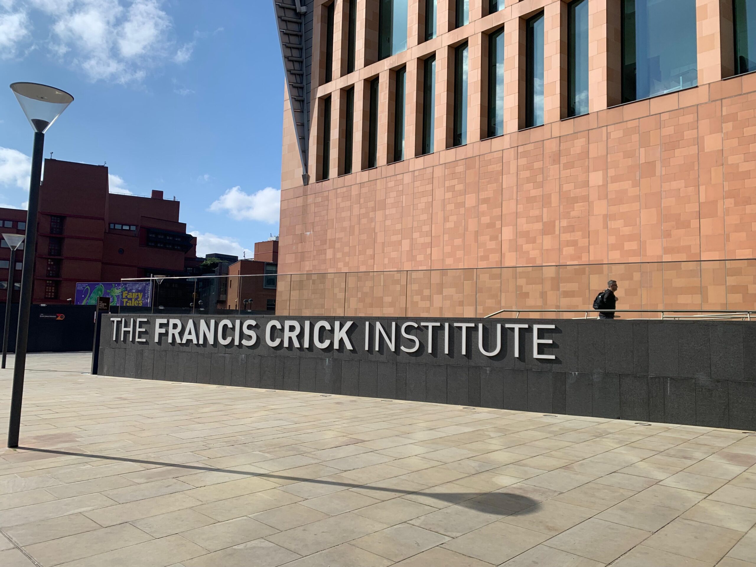
We had the pleasure of interviewing Dr Kurt I. Anderson and Dr. Matt Renshaw from The Francis Crick Institute in London, United Kingdom, and learning more about their imaging research at the Advanced Light Microscopy Science and Technology Platform (CALM STP).
What is your research about?
We are a core facility that supports a wide variety of science relating to development, cancer, and neurobiology, among other areas.
What is your research objective?
We can define two groups of researchers according to their imaging needs. One group wants to image large 3-dimensional samples such as spheroids, organoids, and tissues. For these researchers, the interest is primarily in the location and organisation of cells, often in conjunction with time-lapse imaging to study cellular and tissue dynamics.
The main use has been to image the actin cytoskeleton reorganisation and mitochondrial dynamics in differentiated myotubes in cancer-derived conditioned media and also in pancreatic cancer spheroids. They have also imaged stained blood vessels in fixed whole-mount adipose tissue from mice with cancer cachexia. Projects in their very early stages include expanded samples and also live cells growing in a 3D matrix (e.g., Matrigel).
The second group wants to image the cellular and sub-cellular dynamics of more conventional samples on coverslips. For the second group, a little bit of optical sectioning to improve contrast and high sensitivity to improve sample viability are important. Examples here would be looking for defects in chromosome segregation during mitosis, and mitochondrial and ER dynamics in response to stress conditions in neuronal cells.
How important is imaging in your research?
Imaging is extremely important because of the unique information it provides about biological structure, dynamics, and (through the use of biosensors) biochemistry and signalling. Imaging, especially live imaging, is an essential method, even though it is increasingly only one source of information in complex studies involving other technologies such as metabolomics, transcriptomics, flow cytometry, and computational simulation.
What are the essential characteristics of live cell imaging to you?
Sensitivity, speed, and contrast are probably the three most important aspects of confocal microscopy, along with the ability to multiplex 2-3 colors in a live imaging experiment or 3-5 colors in a fixed sample. Sensitivity is important for enabling fast acquisition with low illumination intensity. This maximises sample longevity and minimises photobleaching during 2D or especially 3D acquisition, which is important whether the goal is short acquisitions with high time resolution or longer acquisitions with lower time resolution. Good contrast is primarily important for resolving the features of thicker samples, but can also be important in the analysis of simple samples like cells in culture.
Which are the characteristics of the NL5+ that you appreciate the most?
We tested the NL5+ extensively and were attracted by two main features: image contrast for the segmentation of cells and nuclei in thick scattering samples like spheroids, organoids, and embryos, and overall sensitivity for live imaging of both thick samples and thin samples, such as imaging mitochondrial dynamics with high time resolution in cells on glass coverslips.
Matt Renshaw – How did the NL5+ help you accelerate your research?
From the perspective of a core facility, I’d say that the challenge is to ensure that we have the right technologies available for when the research groups need them – so that we don’t delay the research! The concept of horizon scanning is to evaluate new and emerging technologies, try to connect them with the plans of the research groups, and then get the right equipment in with limited budgets and space. As more 3D models are being used, we need to make sure we have the right tools to image them. The NL5+ was identified during a horizon scanning exercise when we were interested in knowing more about it and how it could be useful, rather than having a specific project in mind. This led to us identifying a need, and we were able to secure funding for a new system. So, when users approach us with 3D samples we have the NL5+ as an option to complement light-sheet and other confocal approaches.
We would like to thank Dr. Kurt I. Anderson and Dr. Matt Renshaw for this insightful interview.