“Sprouting angiogenesis, the growth of new blood vessels from existing ones, plays a key role in development, wound healing, cardiovascular disease and cancer.” In this webinar, Maxime Cammeraat from the Angiogenesis Laboratory at Amsterdam UMC shows how they image this highly dynamic 3D process in living cells.
Topics covered:
- A high-throughput bead sprouting assay with endothelial cells (HUVECs) in fibrin gels, and automated analysis of sprout number and length.
- Why fixed samples miss key events such as sprout elongation and retraction.
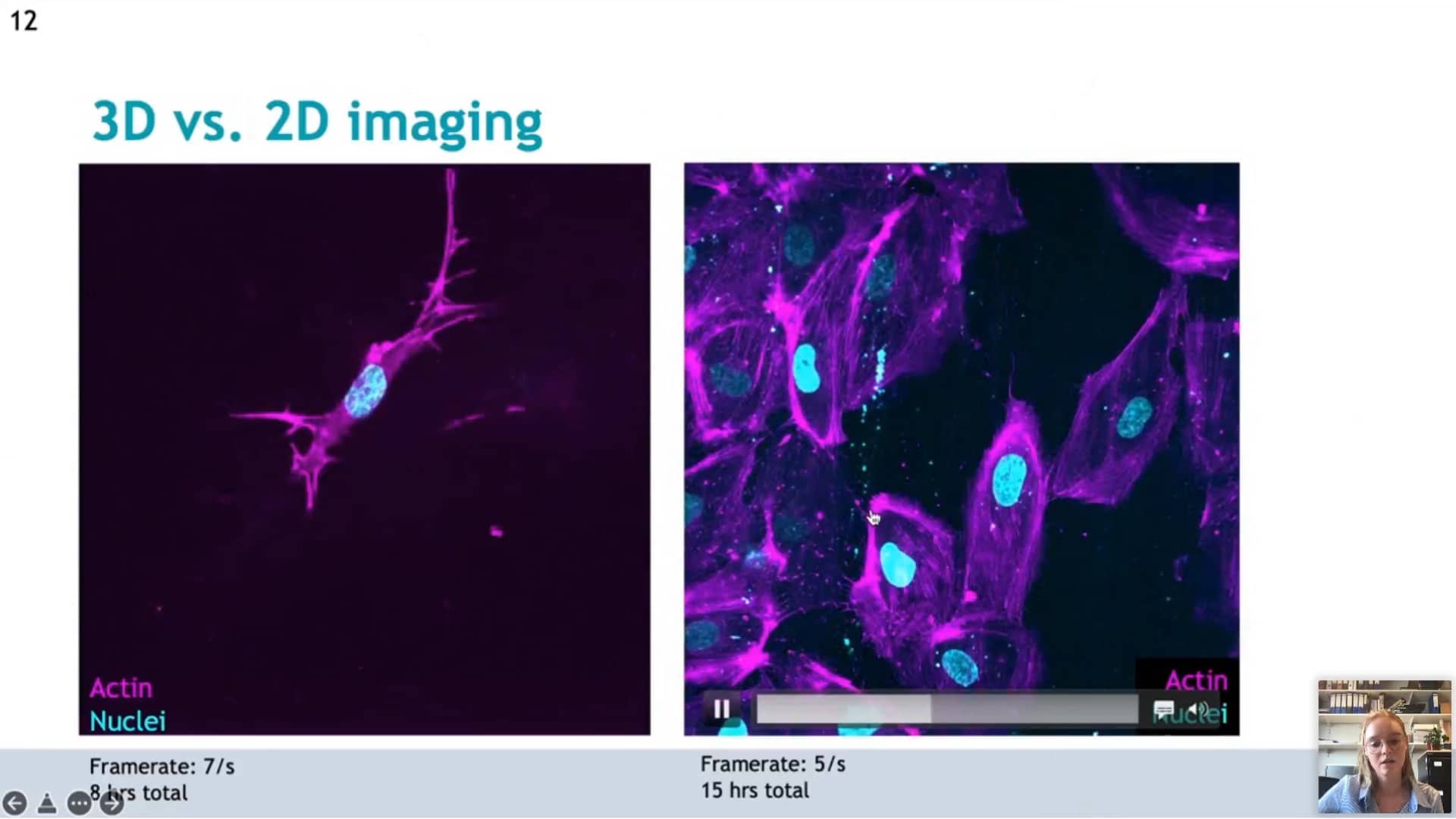
- Non-toxic fluorogenic actin and DNA probes that stay in the medium during imaging, for 2D and 3D time-lapse of up to 15 hours.
- Combining live imaging with RNA interference via lentiviral transduction.
Confocal NL then presents the NL5+ Line REscan confocal and demonstrates it on the sprouting samples, comparing widefield with confocal 3D imaging.