



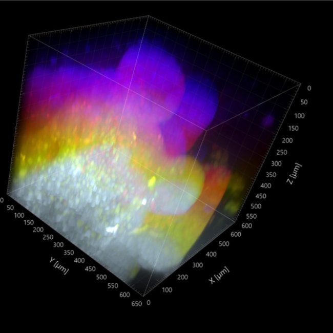
Organoids are physiologically relevant models for studying disease, but their dense 3D architecture makes deep, high-resolution imaging difficult. Expansion microscopy (ExM) physically enlarges the sample, improving resolution, antibody access and optical clarity. In this application note, Visualise.bio and Confocal NL combine ExM with the AION Line REscan confocal, featuring a switchable pinhole, fast large field-of-view imaging and minimal phototoxicity. Expanded mouse small intestinal organoids were imaged more than 600 μm deep in Super resolution, capturing a volume of about 0.3 mm³ in under three minutes without stitching. Combined with automated sample preparation and deformation correction, this workflow offers a robust, scalable platform for deep tissue imaging in organoid research.
Download the application note
Learn more about this application.