
How large is the area that your confocal microscope really images? In this sixth Quick Knowledge, we explain the field of view (FOV) in the sample and on the detector, and why pixels alone make a poor measure of it in REscan confocal microscopy. We show how the Field Number (FN) and the objective magnification give the FOV in units of length, and then compare how RCM2 and GAIA use the camera plane, so you can see why GAIA images a larger FOV with the same camera.

Quick Knowledge Part 6

FIELD OF VIEW
IN RESCAN
CONFOCAL
MICROSCOPY

Field Of View in
Confocal Microscopy

Field Of View (FOV) describes the
area in the sample that a microscope
can image. This area is also known
as FOVsample.

Additionally, FOV represents the portion of the
object that fills the sensor of a detector (FOVdetector).

In standard confocal microscopy FOVsample is usually
equal to FOVdetector and is often expressed in pixels.

Objective

Sample

Tube lens

Scan lens

Laser

Pinhole

Detector

Scanner

Field Of View in
REscan

In REscan confocal, after the first
scan of the sample, the image is
rescanned onto a camera which
works as a detector.

Here, two issues arise if the FOV is expressed in pixels.

Objective

Tube lens

Scan lens

Laser

Pinhole

Scanner

Sample

REscanner

REscan unit

REscan
lens

Detector
(Camera)

Issue 1:
Pixel size varies

Different cameras might have pixel
grids with the same number of pixels,
but different pixel sizes.

2.6 µm per px

4.6 µm per px

6.5 µm per px

12x12 px

12x12 px

12x12 px

Issue 2:
FOVdetector ≠ FOVsample

In REscan confocal, the size of the
camera plane is not always identical
to the FOV in the sample.

FOV can also be expressed as the Field Number
(FN) in the image plane in mm.

Objective

Tube lens

Scan lens

Laser

Pinhole

Scanner

Sample

FN (image plane)

REscanner

REscan unit

REscan
lens

Detector
(Camera)

FOVdetector (pixels in
the camera plane)

FOVsample (sample
plane)

Equation: FOV sample = FN divided by M (Field Number divided by objective magnification)

The solution:
FOV in units of length

The FOVsample is the Field Number
divided by the objective magnification (M).

Different REscan systems handle FOVsample in distinct
ways. For example, this table applies to GAIA
but not to RCM2.

FN

FOV in mm²

Number of
6.5 µm pixels

Number of
4.6 µm pixels

12.5

8.9 x 8.9

1369 x 1369

1935 x 1935

18.8

13.3 x 13.3

2048 x 2048

2898 x 2898

22

15.6 x 15.6

2400 x 2400

3392 x 3392

25

17.7 x 17.7

2728 x 2728

3855 x 3855
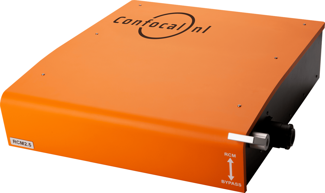
REscan in RCM2

In RCM2, the REscan
amplitude is larger than the
scan amplitude, creating a
super resolution image, yet
limiting FOVsample.

Here, the image plane is
limited to FN12.5 even
though the camera plane is
FN18.8.

Scan on sample

Scan amplitude

REscan on camera

REscan amplitude

FOV in RCM2

An 89 µm by 89 µm
surface on the sample
plane is scanned, when a
100x objective is used.

This is rescanned onto
2048 by 2048 pixels on
the camera with a pixel
size of 6.5 µm.

Scan on sample

Scan amplitude

REscan on camera

REscan amplitude

REscan in GAIA

In GAIA, scan and
REscan amplitude are
always identical,
creating super
resolution images by
using the complete FN
18.8 of the microscope.

The rescanned spot
on the camera is
smaller than the
scanned spot on the
sample.

Scan on sample

Scan amplitude

REscan on camera

REscan amplitude

FOV in GAIA

A 133 µm by 133 µm
surface on the sample
plane is scanned,
when a 100x objective
is used.

This is rescanned onto
2048 by 2048 pixels on
the camera, the same
FOVdetector yet larger
FOVsample when
compared to RCM2.

Scan on sample

Scan amplitude

REscan on camera

REscan amplitude

RCM Point REscan confocal unit GAIA Point REscan super resolution confocal NL5+ Line REscan confocal unit AION Line REscan confocal unit

Point REscan

Line REscan

Download the quick knowledge

Receive more information about this knowledge article.