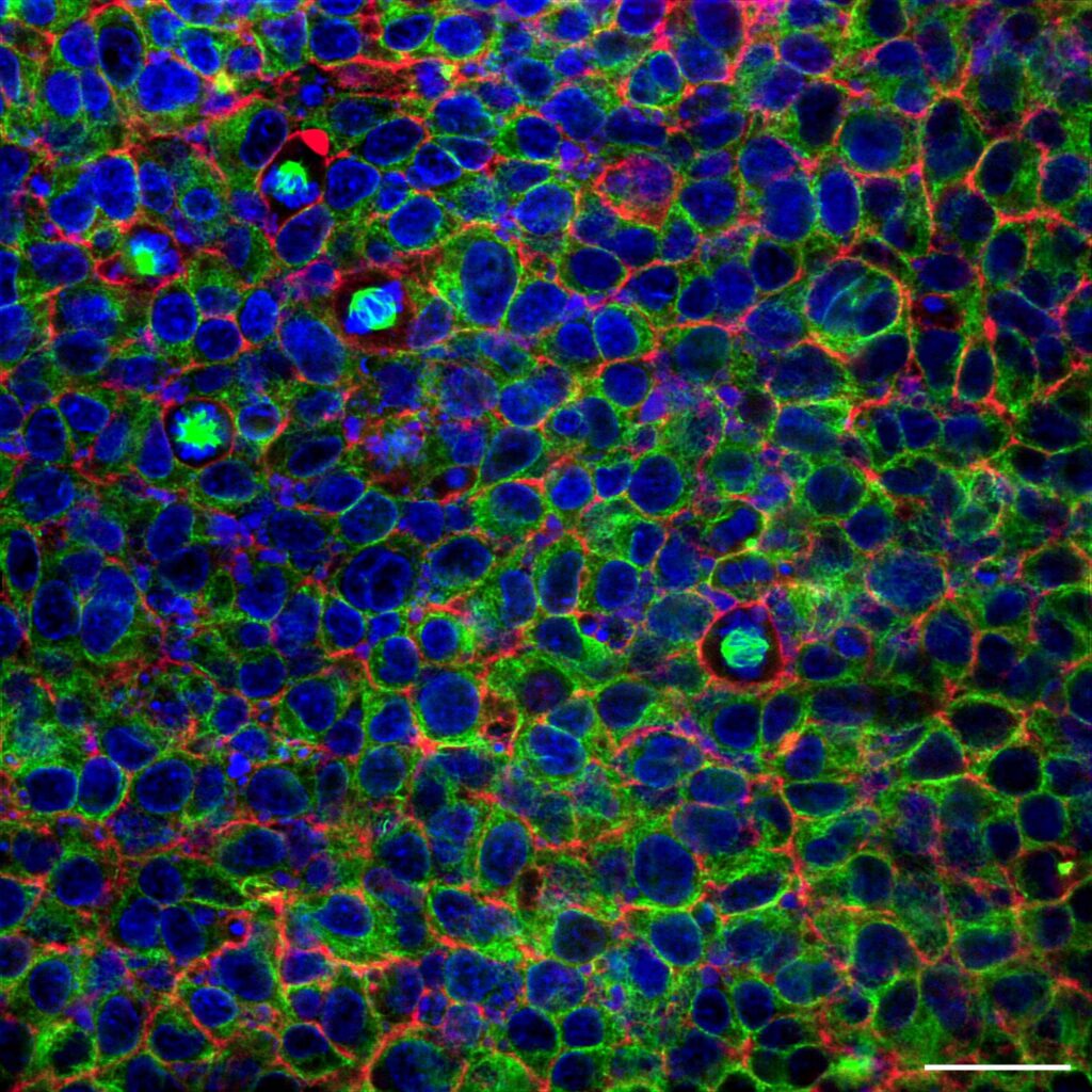
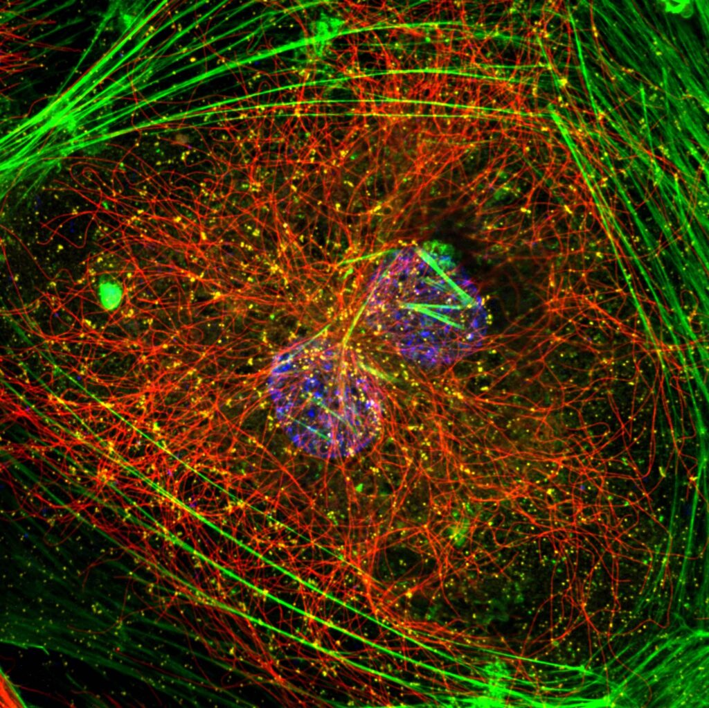
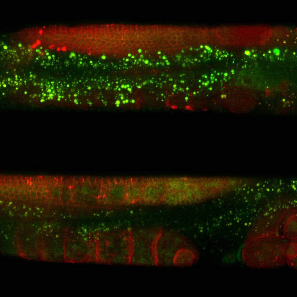
Cell biology focuses on understanding the structure, function, and behaviour of cells. Microscopy is an indispensable tool in this field, providing detailed visualization of cellular components, such as organelles, cytoskeleton, and membranes. Fluorescence microscopy, including confocal, allows the use of labelled markers to track specific proteins or molecules. Advances in super resolution microscopy further enhance spatial resolution, uncovering intricate subcellular details. Together, these techniques illuminate cellular mechanisms, driving discoveries in cell function and drug development.
Explore the applications
Super resolution microscopes that enable deep live cell imaging beyond the diffraction limit using only nanowatts of power.
Developmental biology
Developmental biology investigates how organisms grow and develop, with a focus on cell differentiation, tissue formation, and organogenesis. Zebrafish (Danio rerio) serves as a key model organism due to its transparent embryos and rapid development, allowing real-time observation of developmental processes. Fluorescence and confocal microscopy are commonly used to study labelled proteins, signalling pathways, and organ development at cellular level resolution. Advanced techniques like two-photon and light-sheet microscopy overcome certain limitations of conventional confocal microscopy and allow imaging of live zebrafish embryos in three dimensions, minimizing phototoxicity. Microscopy’s versatility is crucial for understanding the cellular mechanisms underpinning development in zebrafish.
Live cell imaging
Live cell imaging is an approach used to observe and study living cells in real time under a microscope. It involves the use of advanced imaging methods, such as fluorescence, confocal, or time-lapse microscopy, to visualize dynamic cellular processes, including cell division, migration, intracellular trafficking, and signalling.
Live cell imaging is invaluable for understanding cellular behaviour in physiological and pathological contexts, offering insights into biology, disease progression, and the effects of drugs or environmental changes on cellular functions. It prioritizes maintaining cell health and minimizing phototoxicity and photobleaching during observations.
Deep Live cell imaging in Super Resolution
Super resolution (SR) microscopy techniques surpass the diffraction limit, achieving resolutions below 200 nm. These methods utilize advanced strategies like fluorescent molecule localization or selective depletion of excited states to enhance spatial resolution. Super-resolution microscopy is widely applied in cellular and molecular biology to observe subcellular structures, protein interactions, and dynamic processes at a nanoscale level. These techniques have revolutionized our understanding of cellular organization, enabling insights into the molecular mechanisms underlying health and disease.
Neurobiology
In neurobiology, various light microscopy techniques are essential for exploring the complex structures and functions of neurons and neural networks. Confocal microscopy provides high resolution imaging of neuron morphology and synaptic connections in fine detail, especially within brain slices and cultured neurons. Super-resolution techniques like STED (stimulated emission depletion) and SMLM (single molecule localization microscopy like STORM, PALM, PAINT, etc) surpass diffraction limits, revealing nanoscale details in synaptic structures and protein localization. These microscopy techniques collectively advance the understanding of neurobiological processes underlying cognition, behaviour, and disease mechanisms.
Plant Biology
Different types of microscopy are used in plant biology research. To acquire detailed imaging of plant cells, tissues, and organelles with high resolution and specificity, fluorescent microscopy is often chosen. By using fluorescent dyes or autofluorescence, researchers can observe cell walls, chloroplasts, and other structures in living or fixed samples. This technique is essential for studying cellular processes like photosynthesis, nutrient transport, and cell division. Confocal microscopy allows visualization of complex three-dimensional structures, such as root tips or leaf tissues, and tracking of dynamic processes in response to environmental changes.
Ca²⁺ imaging
Calcium imaging refers to the precise visualization of calcium ions (Ca2+) within cells, which play a key role in various signalling pathways. By using calcium-sensitive fluorescent dyes or genetically encoded calcium indicators, microscopy can monitor real-time changes in intracellular calcium concentrations. This is especially valuable in neuroscience and cardiac research, where calcium signalling drives critical functions like neuron firing and muscle contraction.
C. elegans
Microscopy is pivotal in research of the worm Caenorhabditis elegans, enabling the visualization of its transparent body and cellular structures. Researchers commonly use brightfield and differential interference contrast (DIC) microscopy to observe its development, behaviour, and anatomy in vivo. Fluorescence microscopy allows detailed imaging of specific cells, tissues, or molecular processes using fluorescent markers. Advanced techniques like confocal microscopy and two-photon microscopy enable three-dimensional visualization of neural networks, intracellular events, and gene expression patterns. Super resolution microscopy reveals nanoscale structures like synapses or protein complexes. These methods are instrumental in studying key biological phenomena, including neural development, aging, and gene function. Additionally, non-invasive live imaging of C. elegans ensures real-time observation of dynamic processes, making microscopy essential for unravelling its biology.
Organoids
Organoids are three-dimensional, miniature tissue cultures grown from stem cells that replicate the structure and function of real organs, making them essential in biomedical research. They provide an invaluable platform for studying organ development, disease modelling, cancer mechanisms, and drug testing in a way that closely mimics human biology. Unlike traditional cell cultures, organoids contain diverse cell types and exhibit tissue architecture, allowing researchers to observe cellular interactions and responses in a realistic environment. Organoids are also crucial for personalized medicine; patient-derived organoids enable testing treatments tailored to individual genetic profiles, improving therapeutic outcomes. Furthermore, they offer ethical alternatives to animal testing, providing a humane and scalable solution for understanding diseases like cancer, neurological disorders, and infectious diseases.
Cleared tissue
Tissue clearing provides researchers with a tool to investigate intact biological structures in three dimensions. This is achieved by making tissues transparent while preserving their cellular architecture and fluorescence. Tissue clearing removes lipids and other opaque elements, enabling deeper light penetration and clearer imaging. Combined with advanced microscopy techniques like multiphoton or light-sheet microscopy, cleared tissue imaging provides detailed views of complex systems, such as neural networks or vascular structures, within whole organs or large tissue sections. It is especially valuable in neuroscience, developmental biology, and pathology, where understanding spatial relationships and large-scale structures is crucial.
Satisfied researchers
around the globe
Our latest news
Subscribe to our newsletter
Only sharing updates that you can’t miss. Stay up to date with our latest application notes, publications, webinars, news & events and important company updates!


