


Gabor Steinbach, David Nagy, Gábor Sipka, Erik Manders, Győző Garab, László Zimányi | Eur Biophys J. 2019 Jul;48(5):457-463. doi: 10.1007/s00249-019-01365-4. Epub 2019 Apr 13
Summary
Confocal laser scanning microscopy is a widely used imaging technique in biology, medicine, and materials science for visualizing complex cellular structures and molecular assemblies. Recent advances have improved system performance through optical upgrades while maintaining usability and cost efficiency.
Differential polarization (DP) attachments enable precise 2D and 3D mapping of sample anisotropy alongside standard fluorescence or transmission imaging. Re-scan confocal microscopy (RCM) has also been shown to improve resolution, signal-to-noise ratio, and image quality.
In this study, the authors combined RCM with a DP attachment using a liquid crystal retarder synchronized with image acquisition. With dedicated software, the system enabled simultaneous fluorescence-detected linear dichroism (FDLD) and confocal fluorescence imaging.
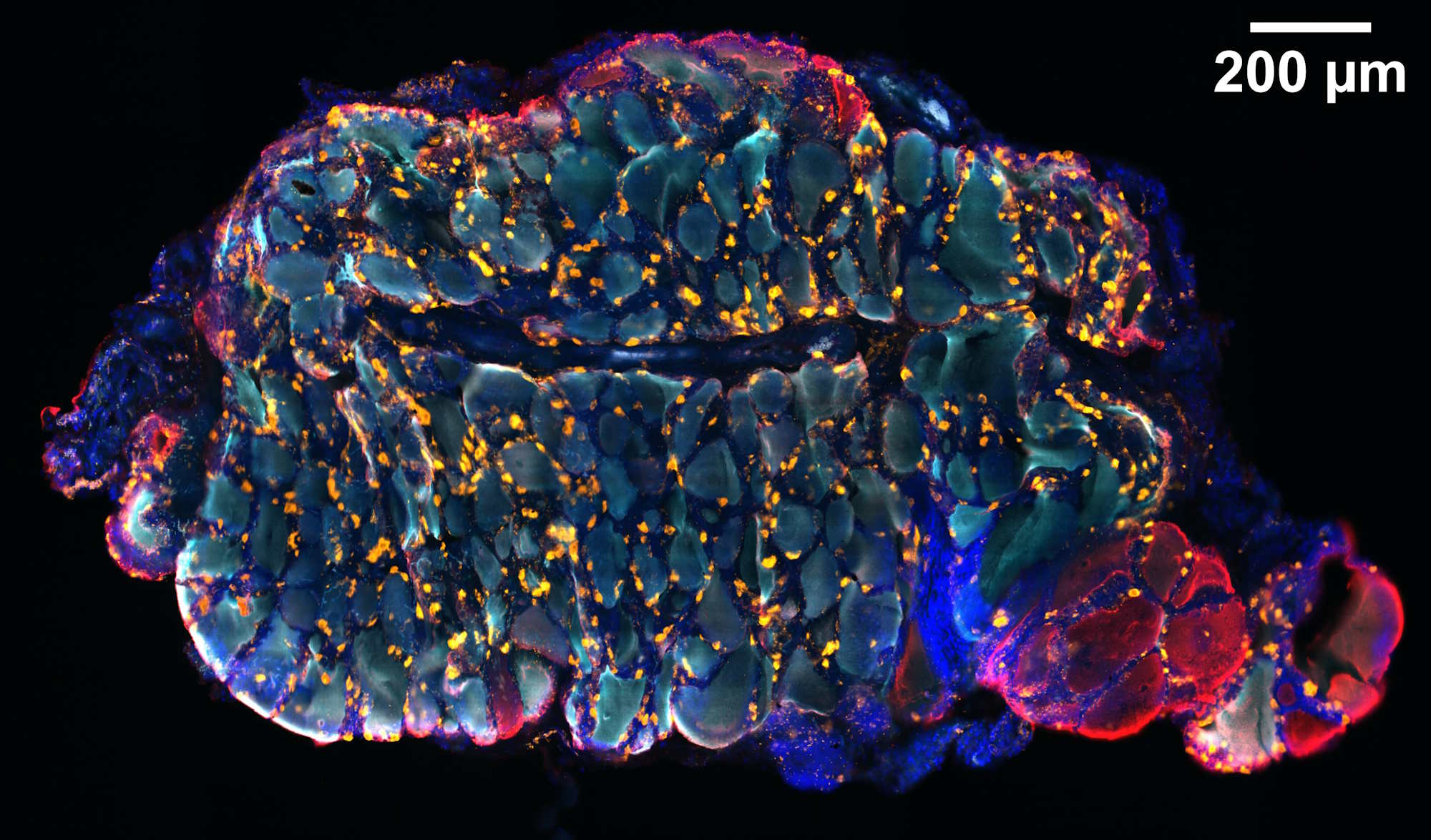
FDLD reveals anisotropic molecular organization beyond conventional intensity imaging. Demonstrated on stained Ginkgo biloba cell walls, the method highlights the potential of combining enhanced resolution with molecular orientation mapping in a single platform.
Read the publication: https://pubmed.ncbi.nlm.nih.gov/30982120