Live cell imaging

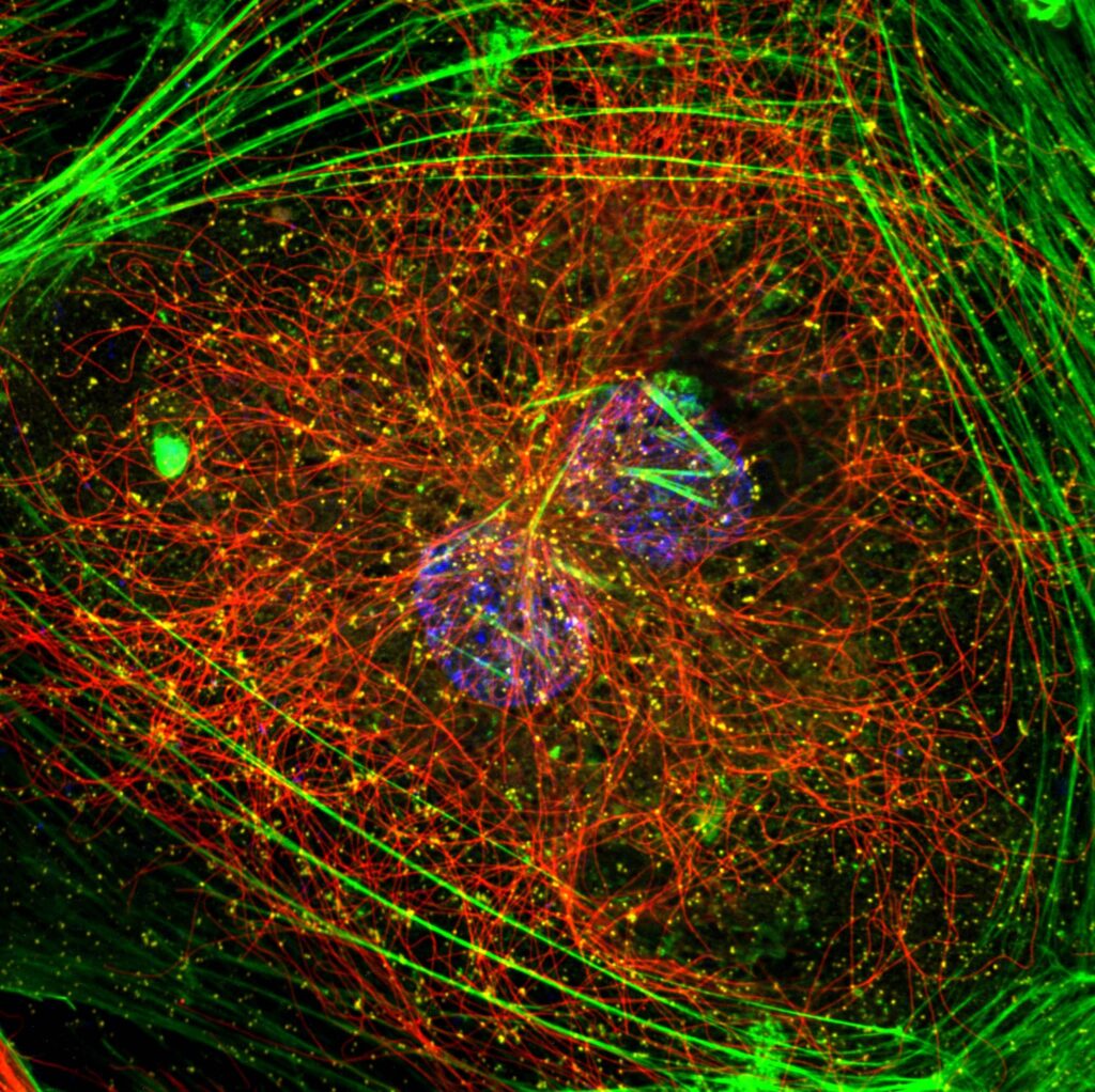
Super resolution microscopes that enable deep live cell imaging beyond the diffraction limit using only nanowatts of power.

Study living cells in real time

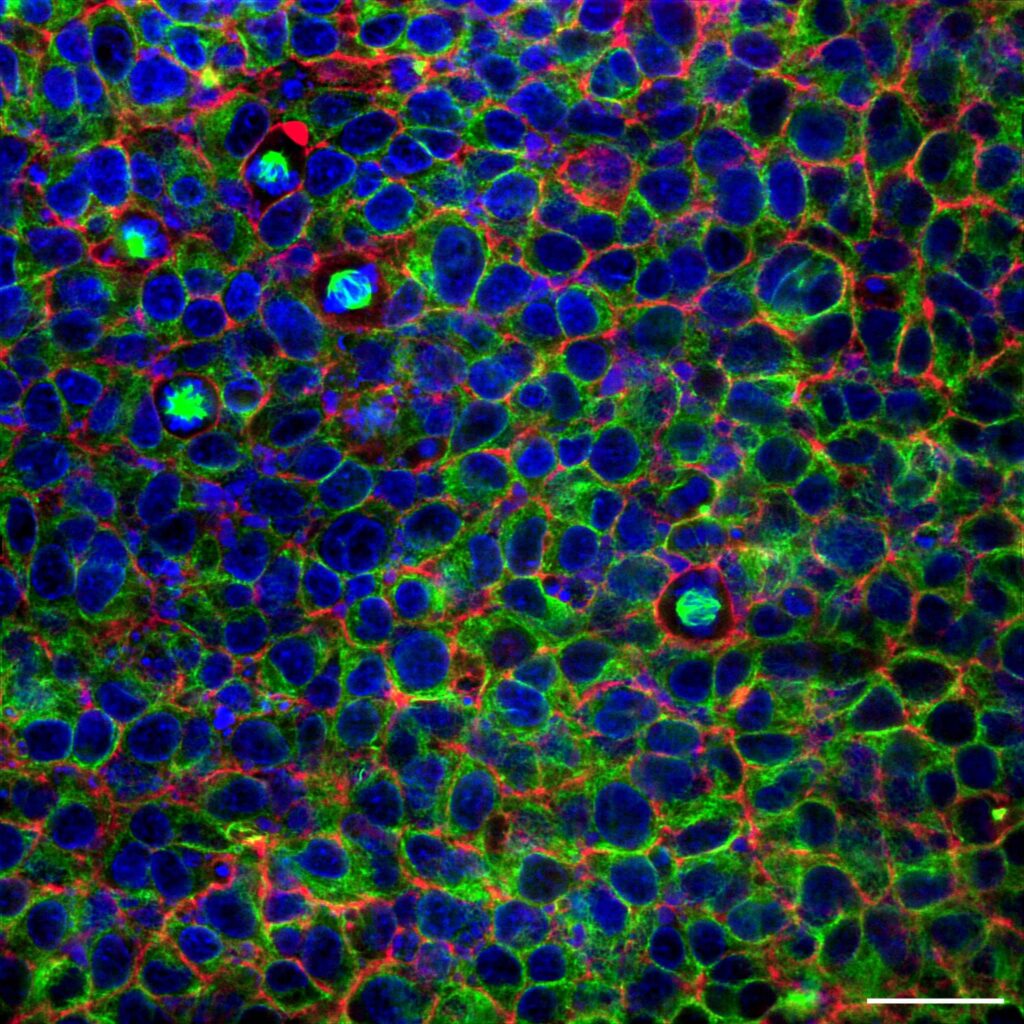
Live cell imaging is an approach used to observe and study living cells in real time under a microscope. It involves the use of advanced imaging methods, such as fluorescence, confocal, or time-lapse microscopy, to visualize dynamic cellular processes, including cell division, migration, intracellular trafficking, and signalling.

Live cell imaging is invaluable for understanding cellular behaviour in physiological and pathological contexts, offering insights into biology, disease progression, and the effects of drugs or environmental changes on cellular functions. It prioritizes maintaining cell health and minimizing phototoxicity and photobleaching during observations.

High-resolution and three-dimensional visualization

For live cell imaging, confocal microscopy allows researchers to track dynamic events like cell division, protein interactions, and intracellular transport, albeit not suitable for observing living cells over extended periods. Phototoxicity is the main adverse factor for the application of confocal microscopy in live cell imaging, as the strong laser light damages cells over prolonged imaging.

Photobleaching is another issue, where fluorescence decays, reducing image quality over time. Additionally, the limited depth of field and slower image acquisition can hinder capturing fast cellular dynamics in thicker tissue samples.

Track dynamic events

For live cell imaging, confocal microscopy allows researchers to track dynamic events like cell division, protein interactions, and intracellular transport, albeit not suitable for observing living cells over extended periods. Phototoxicity is the main adverse factor for the application of confocal microscopy in live cell imaging, as the strong laser light damages cells over prolonged imaging.

Photobleaching is another issue, where fluorescence decays, reducing image quality over time. Additionally, the limited depth of field and slower image acquisition can hinder capturing fast cellular dynamics in thicker tissue samples.

Products

Discover our solutions

Find the appropriate confocal system for your imaging needs.

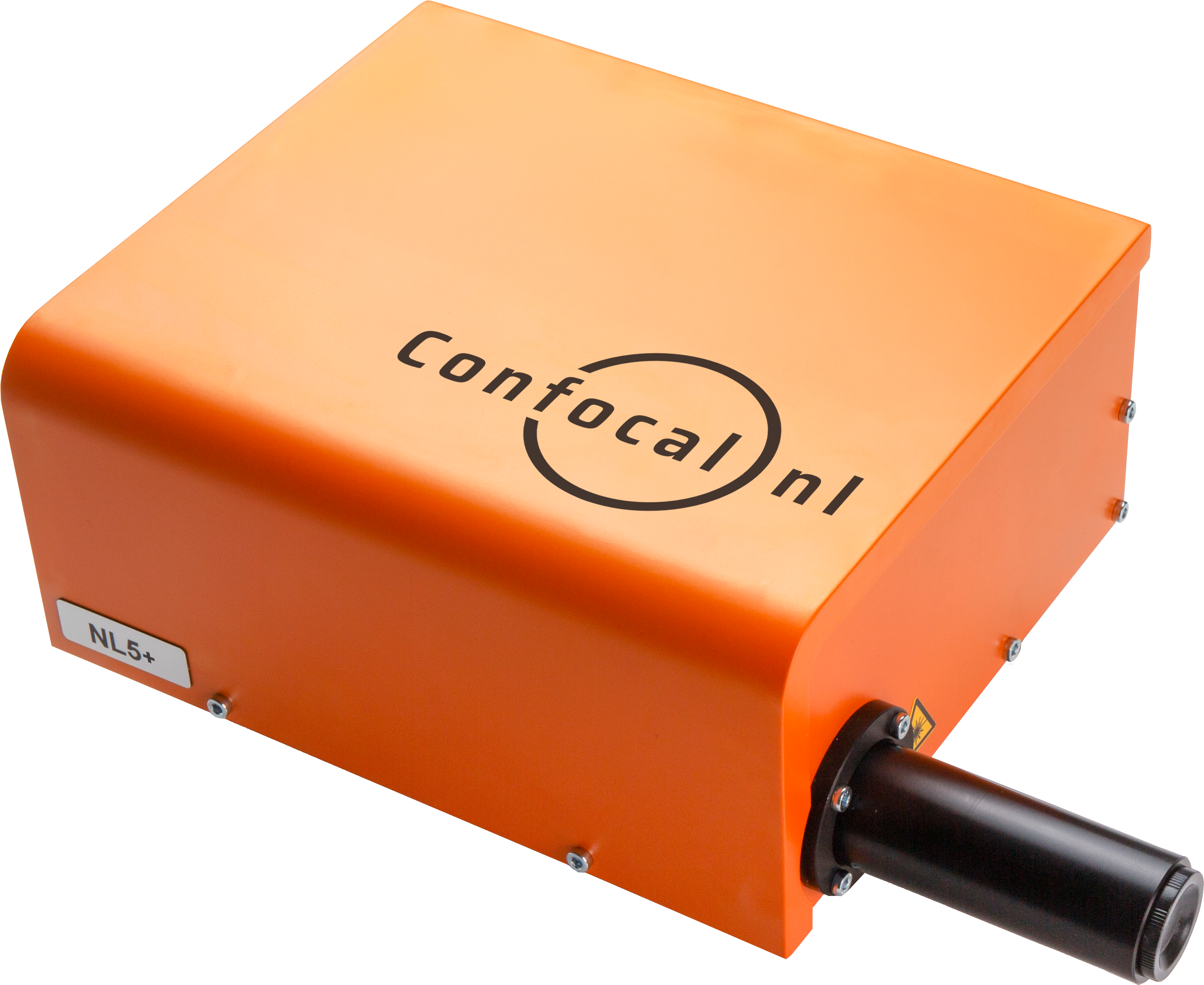
NL5

NL5 is a fast confocal system with high sensitivity and resolution. Quickly screen a multi-well plate with multicolor images, and select the most promising ones.

Learn more

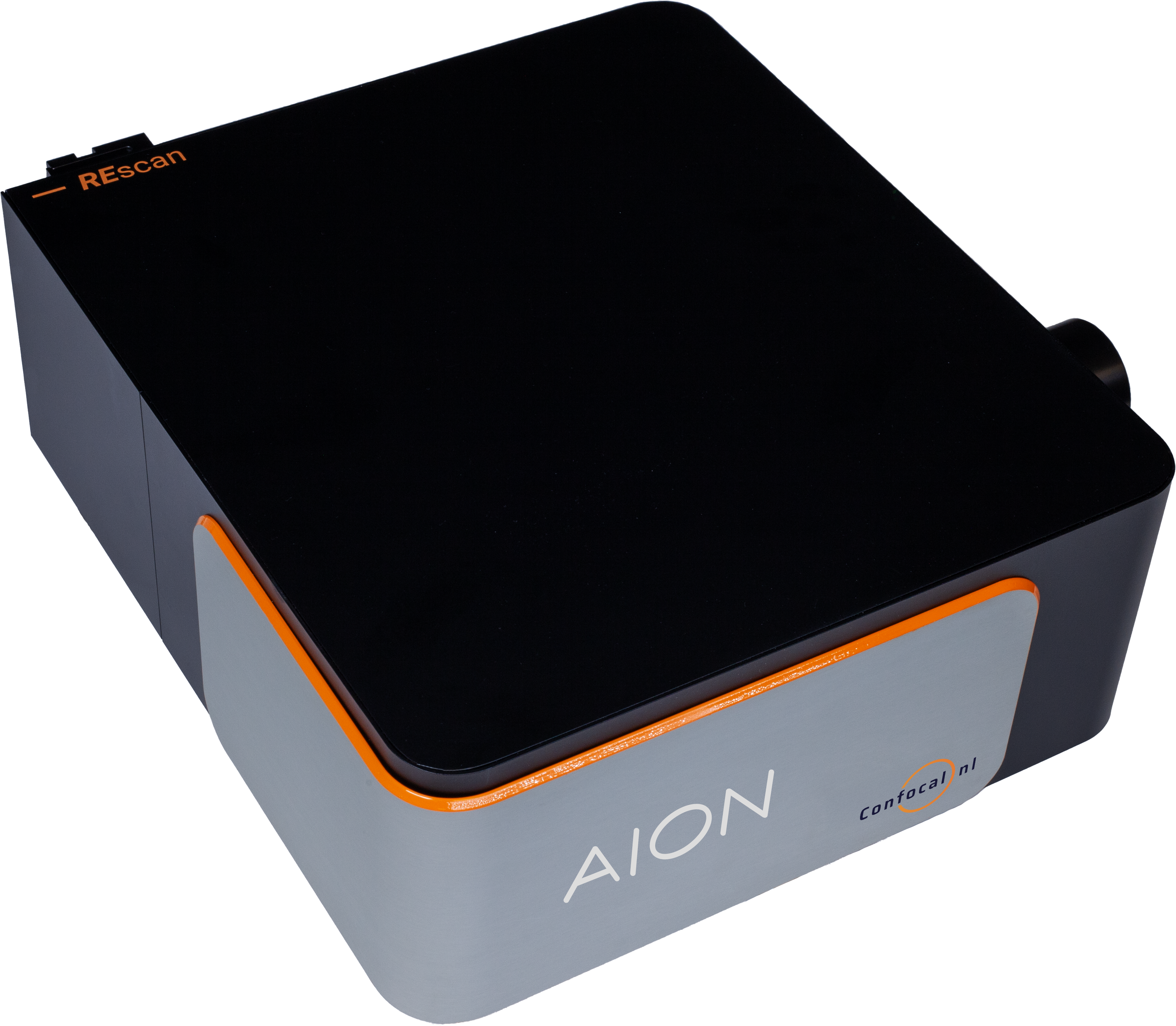
AION

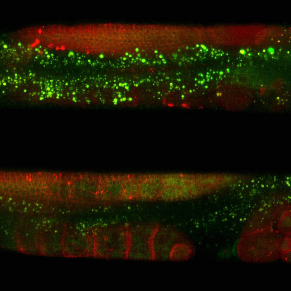
AION is the third generation of our fast confocal technology. It provides high contrast images from thicker specimens such as organoids, tissue samples, plant and animal model organisms.

Learn more

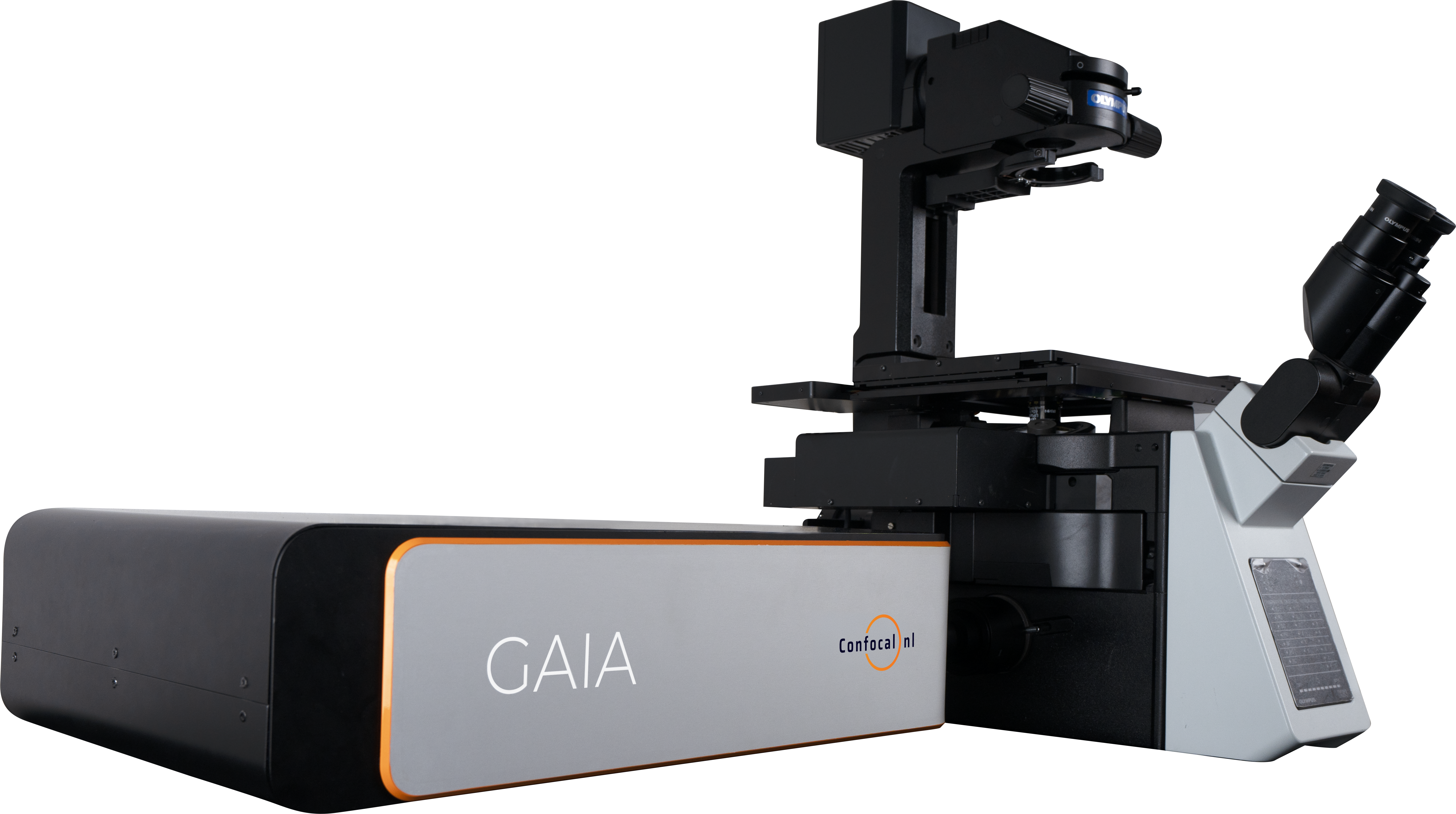
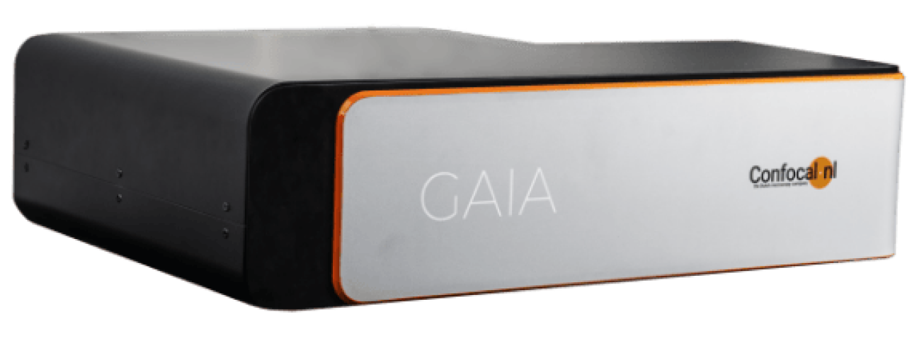
GAIA

The perfect solution for live cell super resolution imaging thanks to its very low phototoxicity.

Learn more

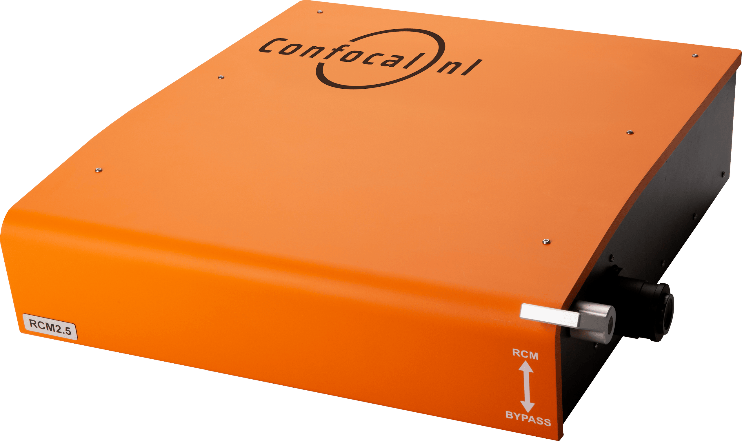
RCM

Capture datasets with 170nm raw resolution (120nm after deconvolution) by using 60x to 100x high numerical aperture (NA) magnification objectives and keep the laser intensity at a minimum.

Learn more
RCM Confocal NL | Discover super resolution imaging with Re-scan Confocal Microscope

NL5

NL5

NL5 is a fast confocal system with high sensitivity and resolution. Quickly screen a multi-well plate with multicolor images, and select the most promising ones.

Learn more

AION

AION

AION is the third generation of our fast confocal technology. It provides high contrast images from thicker specimens such as organoids, tissue samples, plant and animal model organisms.

Learn more

GAIA

GAIA

The perfect solution for live cell super resolution imaging thanks to its very low phototoxicity.

Learn more

RCM

RCM

Capture datasets with 170nm raw resolution (120nm after deconvolution) by using 60x to 100x high numerical aperture (NA) magnification objectives and keep the laser intensity at a minimum.

Learn more
Resources

Related Resources