


Thanks to its patented confocal technology, GAIA Point REscan is a super resolution microscope enabling deep live cell imaging beyond the diffraction limit using only microwatts of power. Our Point REscan is available in two versions, streamlined GAIA α and flagship GAIA λ.
GAIA
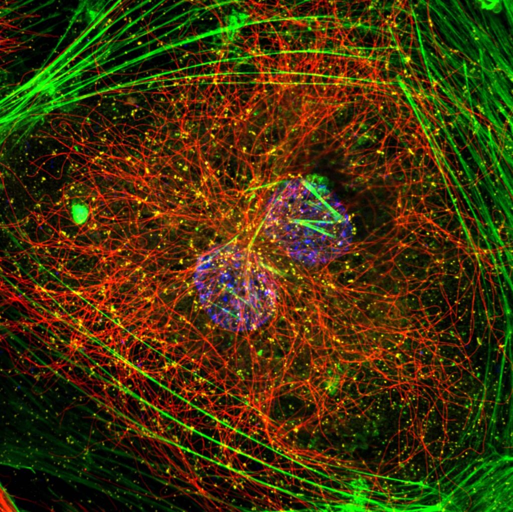
Live cell imaging in Super resolution
Image deep in super resolution
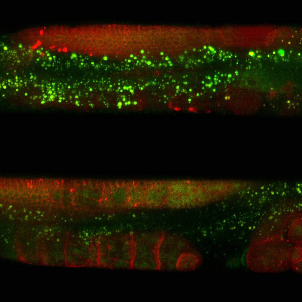
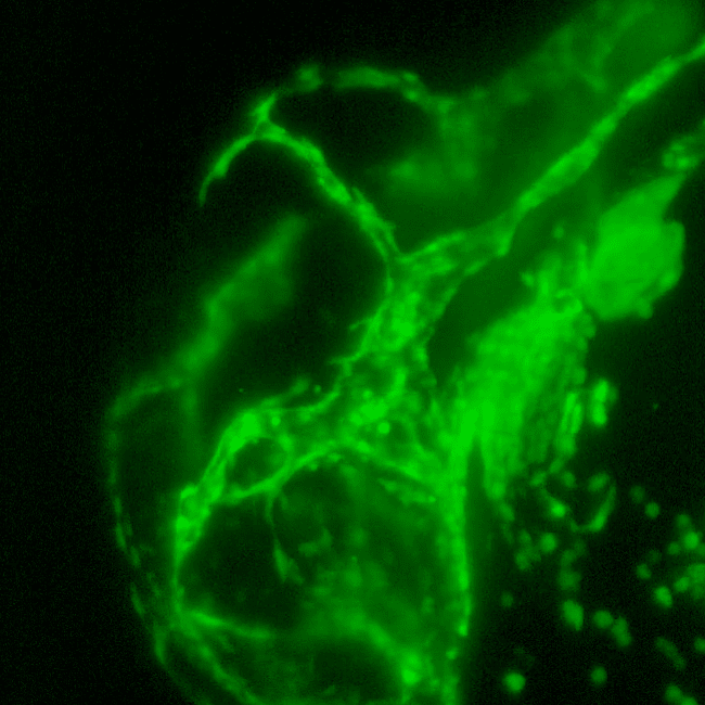
A combination of low laser power requirements, high sensitivity of the detector and a novel optical design present in GAIA Point REscan enables super resolution beyond 500μm of depth. This makes GAIA Point REscan confocal a perfect solution for live cell super resolution imaging of even thicker specimens like cleared tissue, whole zebrafish embryos, organoids and spheroids.
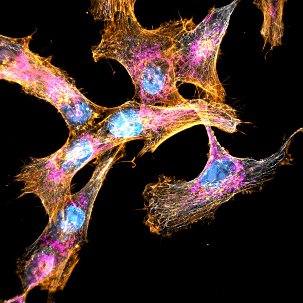
HeLa Cells, Image courtesy of Nicolas Touret (University of Alberta, Edmonton, Canada).
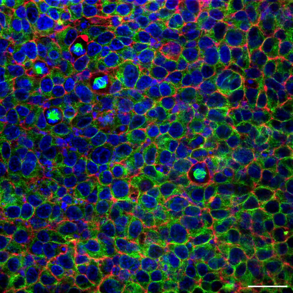
Image wide in super resolution
The disruptive technology in GAIA provides more than double the field of view in the sample plane compared to its predecessor. Consequently, GAIA enables super resolution imaging over a large FOV and using a wide range of objectives (30X -100X). For the best images high NA objectives are a must.
Concept of RCM Point REscan
Instead of using a single detector, we use a second scanner, i.e. REscanner, which is synchronized with the first scanner and REscans the emission light onto a camera.
This way the signal automatically ends up at the right place on the camera and the image is reconstructed all by itself.
Optical redesign in GAIA Point REscan confocal
For GAIA, our revolutionary new Point REscan confocal, we added bespoke optical elements before and after the pinhole. This provides the improvement of resolution beyond the diffraction limit while increasing the effective field of view (FOV).
As an added value, we introduced a motorized switchable pinhole that enables perfect confocality for a wide range of objectives (4X-100X). For proper super resolution, one should use high NA objectives.
Super resolution with Point REscan confocal
In Point REscan confocal, the magnification of the image and the spot are decoupled. Thus, one can magnify the image compared to the spot.
In RCM, we do this by giving the REscanner a larger amplitude than the scanner. As a consequence, all the details in the image are pulled further apart and the image is in super resolution.
However, this does not give infinite improvement in resolution. It turns out that the optimal situation is provided by twice as large REscan amplitudes, which improves the resolution by 1.4x*.
Creating a smaller spot
In GAIA, we took a different approach. Here, we are making the spot smaller instead of the image larger by using bespoke optical elements before and after the pinhole.
At the same time, we are retaining 1.4x resolution improvement.
In addition, we gain more than double the FoV in the sample plane as well as an improvement in speed.
Low phototoxicity of Point REscan confocal
GAIA Point REscan confocal is a perfect solution for live cell super resolution imaging thanks to its very low phototoxicity.
Low phototoxicity is attributed to two important factors:
- GAIA, as well as all members of Point REscan family, has the ability to image using a large pinhole, thereby collecting more light.
- The sensor in Point REscan has a very high sensitivity.
Motorized pinhole switch
A pinhole serves to block the out-of-focus light. In practice, a pinhole size between 1 and 2 Airy units (1-2x times as large as the spot) gives the best images.
If the pinhole is smaller, the signal is sacrificed. If it is larger, confocality is lost.
WIth a motorized pinhole switch, we are ensuring optimal confocality for a wide range of objectives (4x-100X).
Our Point REscans can be mounted on pretty much any microscope body. Adding a camera and a laser gives rise to fully equipped confocal Super resolution microscope. Because it does not need a high laser power, it has a very low phototoxicity, making it perfectly suite for live cell imaging.
Systems
Discover GAIA systems that meet your needs
GAIA α
$ 99
Per MonthDetector
Camera (sCMOS)
Resolution in real time
120 nm deconvolved, 170 nm raw image
Detector sensitivity
Up to 95% QE
FOV
FN18: 330×330 μm in super resolution using 40x objective
Speed in line scanning
3 fps at 512 x 512 px, 30 fps in sprint level max at 256 x 256 px
Wavelength
VIS+NIR (400-1100 nm)
Software
KRONOS, Micromanager, SDK available for integration on request
Deconvolution
SVI Hyugens (post processing)
Modalities
Super resolution, Widefield, Brightfield
Pinhole
Fixed
Emission filter
Single band filter wheel, optional quad band only
Adaptability
All commercially available bodies
GAIA λ
$ 99
Per MonthDetector
Camera (sCMOS)
Resolution in real time
120 nm deconvolved, 170 nm raw image
Detector sensitivity
Up to 95% QE
FOV
FN18: 330×330 μm in super resolution using 40x objective
Speed in line scanning
3 fps at 512 x 512 px, 30 fps in sprint level max at 256 x 256 px
Wavelength
VIS+NIR (400-1100 nm)
Software
KRONOS, Micromanager, SDK available for integration on request
Deconvolution
SVI Hyugens (post processing)
Modalities
Super resolution, Widefield, Brightfield
Pinhole
Switchable
Emission filter
Motorized single band filter wheel
Adaptability
All commercially available bodies
Resources
Related Resources
Download the product flyer
Receive more information on this product or request a quote.