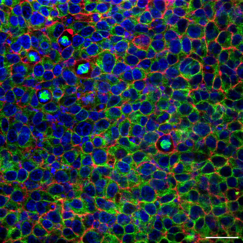
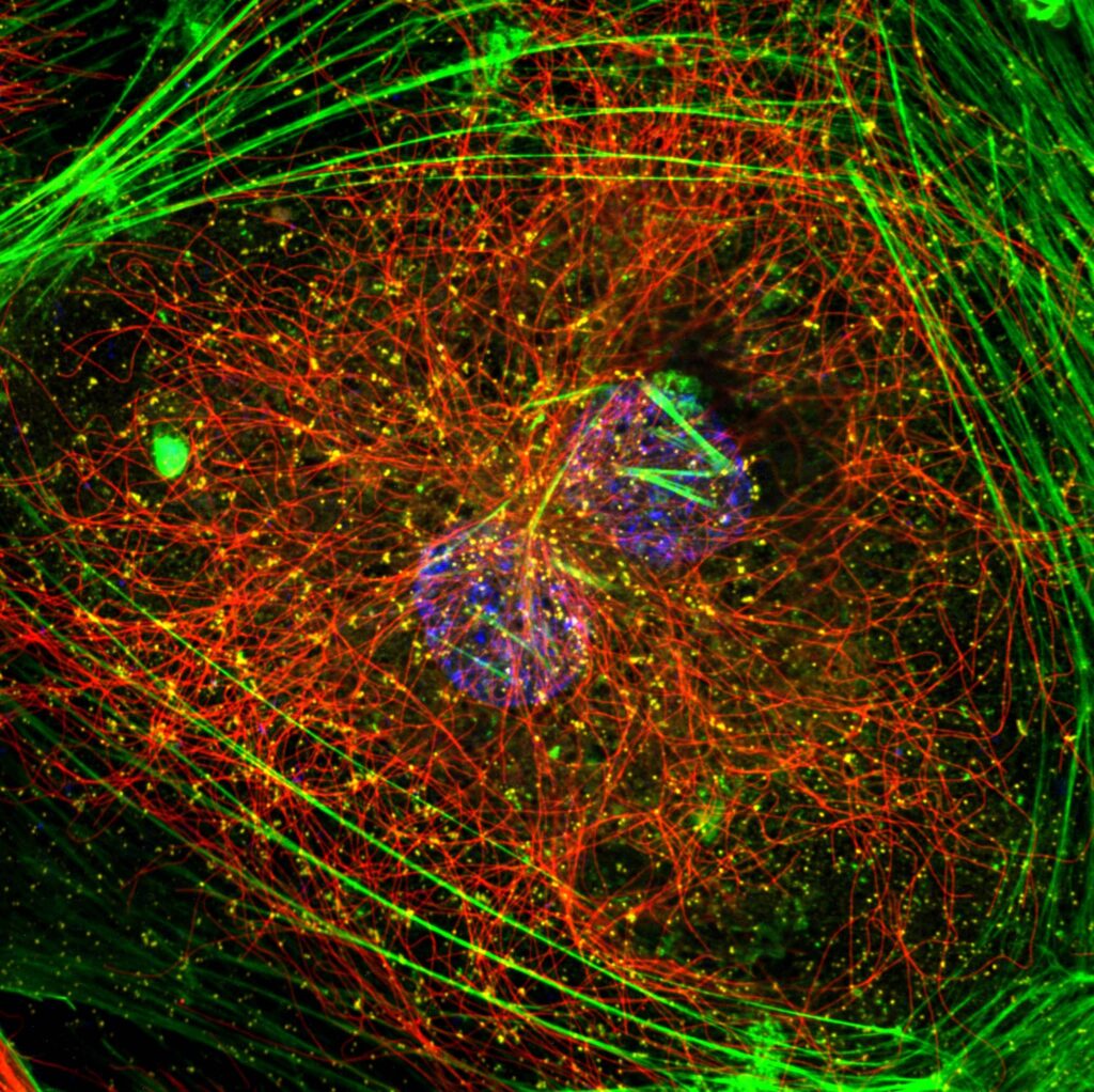
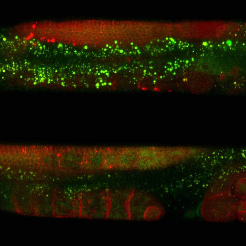
Developmental biology investigates how organisms grow and develop, with a focus on cell differentiation, tissue formation, and organogenesis. Zebrafish (Danio rerio) serves as a key model organism due to its transparent embryos and rapid development, allowing real-time observation of developmental processes. Fluorescence and confocal microscopy are commonly used to study labelled proteins, signalling pathways, and organ development at cellular level resolution. Advanced techniques like two-photon and light-sheet microscopy overcome certain limitations of conventional confocal microscopy and allow imaging of live zebrafish embryos in three dimensions, minimizing phototoxicity. Microscopy’s versatility is crucial for understanding the cellular mechanisms underpinning development in zebrafish.
Developmental biology
Super resolution microscopes that enable deep live cell imaging beyond the diffraction limit using only nanowatts of power.
Investigates how organisms grow
Increased depth penetration
Confocal microscopy in zebrafish developmental biology faces challenges such as limited depth penetration in thickening embryos, restricting visualization of deeper structures. Prolonged imaging can cause phototoxicity, disrupting normal development. Additionally, the rapid cellular dynamics of organogenesis often exceed the temporal resolution of confocal systems, making it difficult to capture transient events accurately.
Imaging with minimal phototoxicity
Line REscan confocal AION and NL5+ enable imaging of the zebrafish embryos’ development post fertilization over extended periods with minimal phototoxicity. Observing rapid dynamic events during development over a large field of view is made possible through line REscan technology. This technology also enables imaging beyond 500 um in depth, overcoming the obstacles present with conventional confocal microscopy.
Discover our solutions
Find the appropriate confocal system for your imaging needs.
NL5
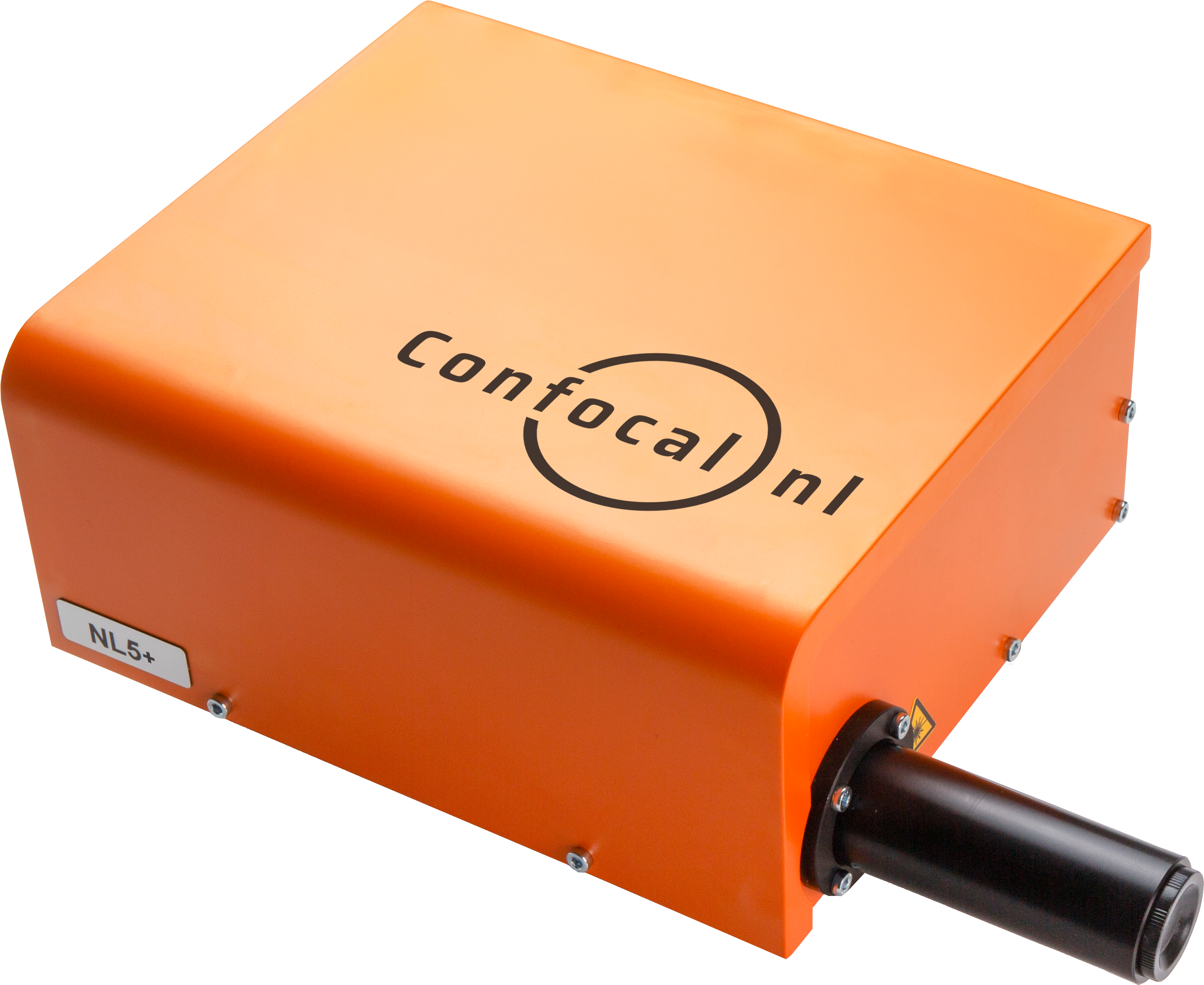
NL5 is a fast confocal system with high sensitivity and resolution. Quickly screen a multi-well plate with multicolor images, and select the most promising ones.
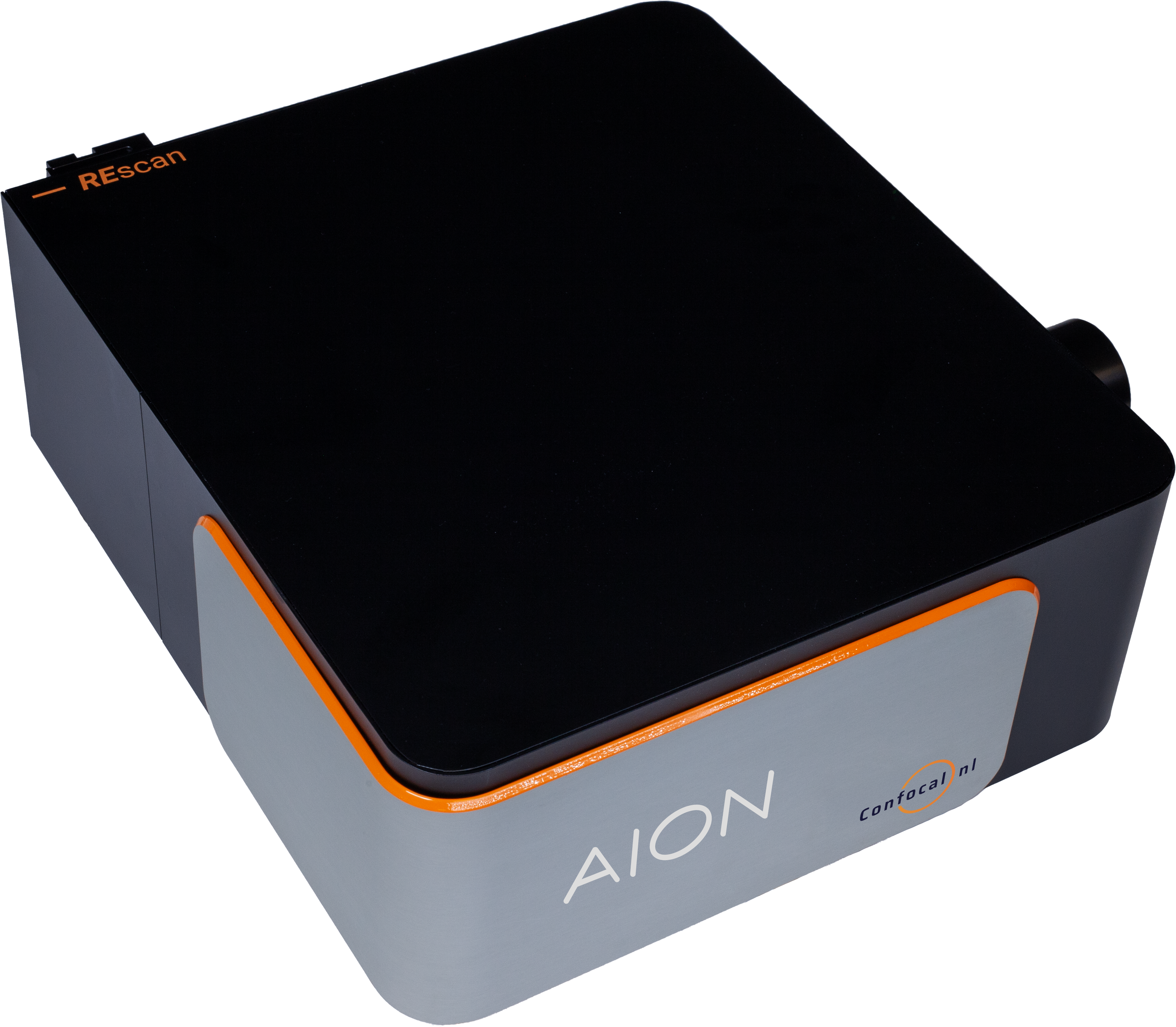
AION
AION is the third generation of our fast confocal technology. It provides high contrast images from thicker specimens such as organoids, tissue samples, plant and animal model organisms.
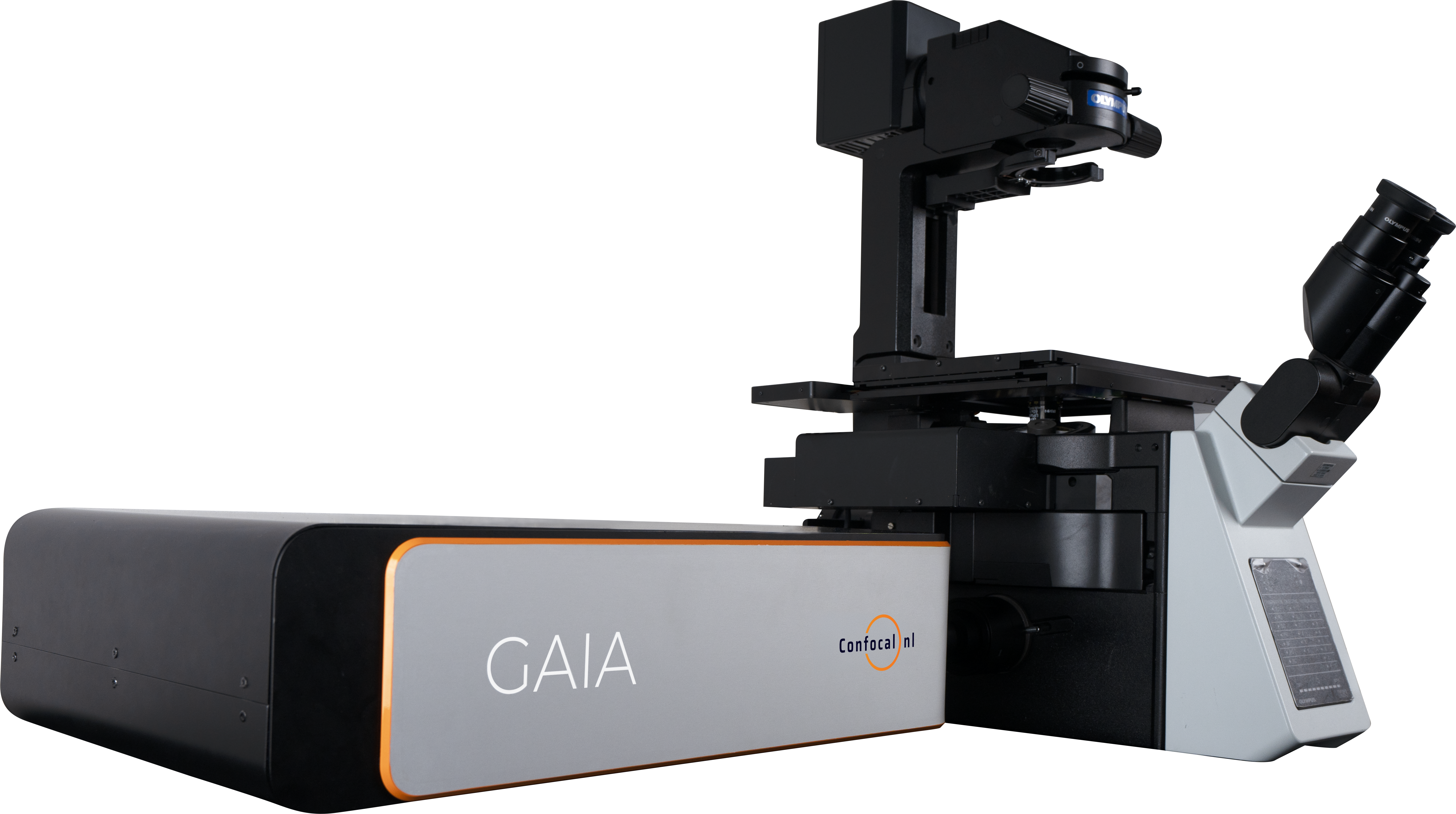
GAIA
The perfect solution for live cell super resolution imaging thanks to its very low phototoxicity.
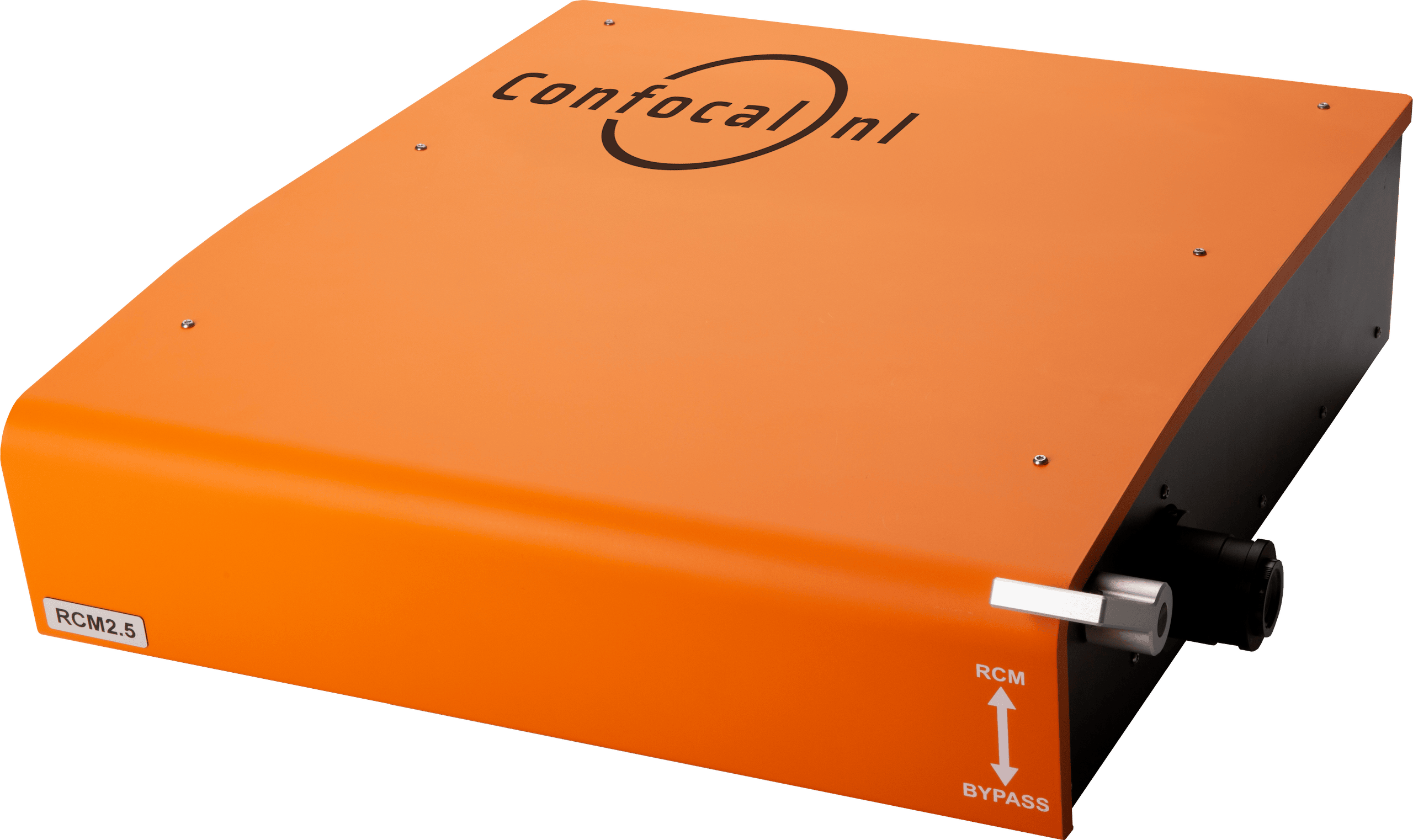
RCM
Capture datasets with 170nm raw resolution (120nm after deconvolution) by using 60x to 100x high numerical aperture (NA) magnification objectives and keep the laser intensity at a minimum.
NL5

NL5
NL5 is a fast confocal system with high sensitivity and resolution. Quickly screen a multi-well plate with multicolor images, and select the most promising ones.
AION

AION
AION is the third generation of our fast confocal technology. It provides high contrast images from thicker specimens such as organoids, tissue samples, plant and animal model organisms.
GAIA

GAIA
The perfect solution for live cell super resolution imaging thanks to its very low phototoxicity.
RCM
RCM
Capture datasets with 170nm raw resolution (120nm after deconvolution) by using 60x to 100x high numerical aperture (NA) magnification objectives and keep the laser intensity at a minimum.


