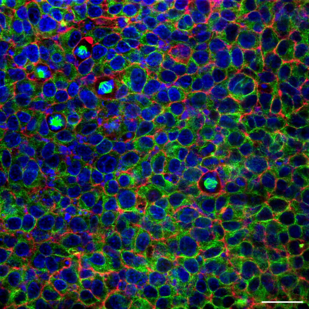
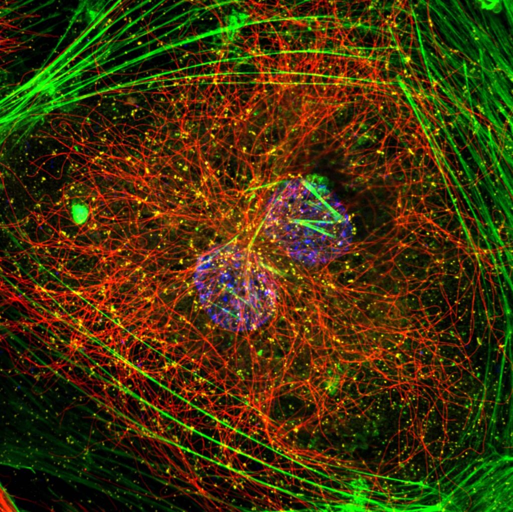
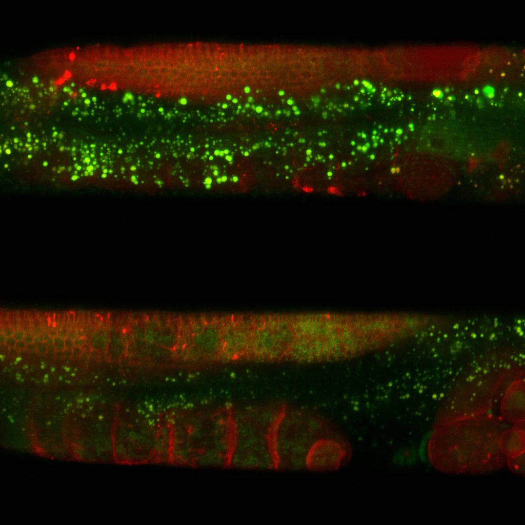
Microscopy is pivotal in research of the worm Caenorhabditis elegans, enabling the visualization of its transparent body and cellular structures. Researchers commonly use brightfield and differential interference contrast (DIC) microscopy to observe its development, behaviour, and anatomy in vivo. Fluorescence microscopy allows detailed imaging of specific cells, tissues, or molecular processes using fluorescent markers. Advanced techniques like confocal microscopy and two-photon microscopy enable three-dimensional visualization of neural networks, intracellular events, and gene expression patterns. Super resolution microscopy reveals nanoscale structures like synapses or protein complexes. These methods are instrumental in studying key biological phenomena, including neural development, aging, and gene function. Additionally, non-invasive live imaging of C. elegans ensures real-time observation of dynamic processes, making microscopy essential for unravelling its biology.
C. elegans
Super resolution microscopes that enable deep live cell imaging beyond the diffraction limit using only nanowatts of power.
Study key biological phenomena of C. elegans
Limit phototoxicity and photobleaching
Confocal microscopy in C. elegans research faces challenges such as limited tissue penetration due to the worm’s thick cuticle and dense tissues. Phototoxicity and photobleaching from prolonged imaging can disrupt normal behaviour and physiology. Additionally, immobilization for live imaging may introduce stress, potentially altering cellular processes and affecting experimental accuracy.
Deep imaging with minimal phototoxicity
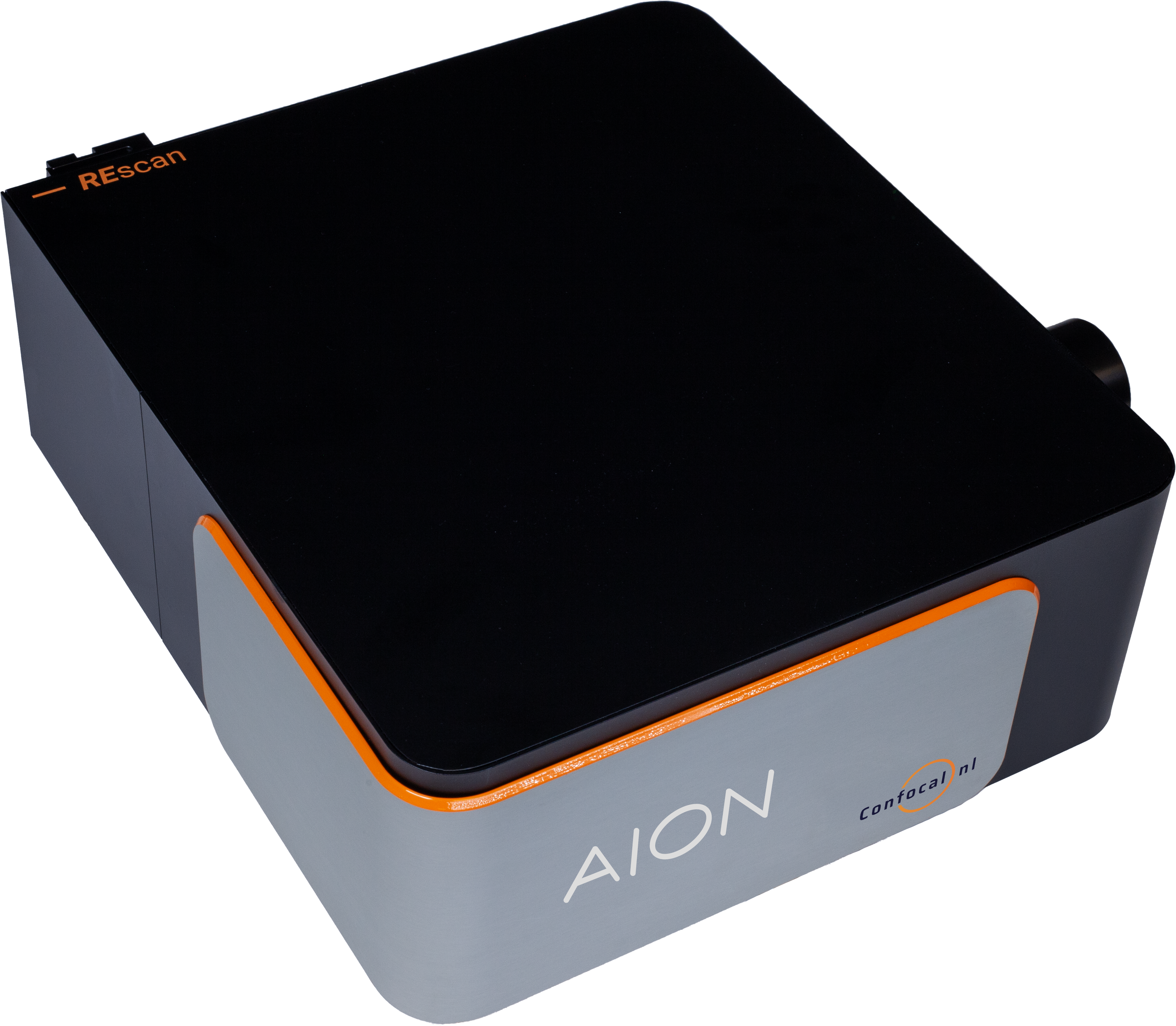
AION enables deep imaging through C. elegans dense tissue with minimal phototoxicity and photobleaching. Due to its speed of over 100 fps, with the largest field of view, it provides detailed structures and dynamic events within the moving worm over prolonged periods.
Discover our solutions
Find the appropriate confocal system for your imaging needs.
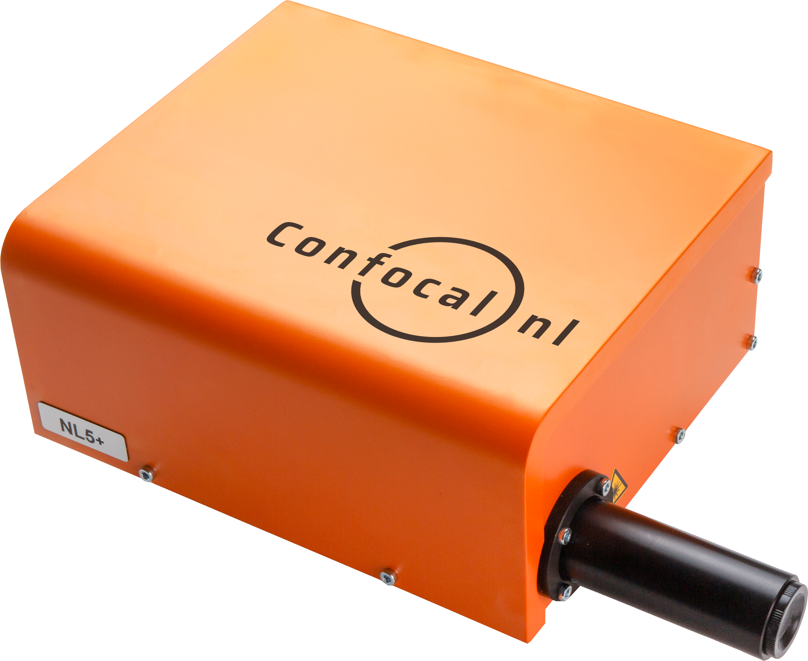
NL5
NL5 is a fast confocal system with high sensitivity and resolution. Quickly screen a multi-well plate with multicolor images, and select the most promising ones.
AION
AION is the third generation of our fast confocal technology. It provides high contrast images from thicker specimens such as organoids, tissue samples, plant and animal model organisms.
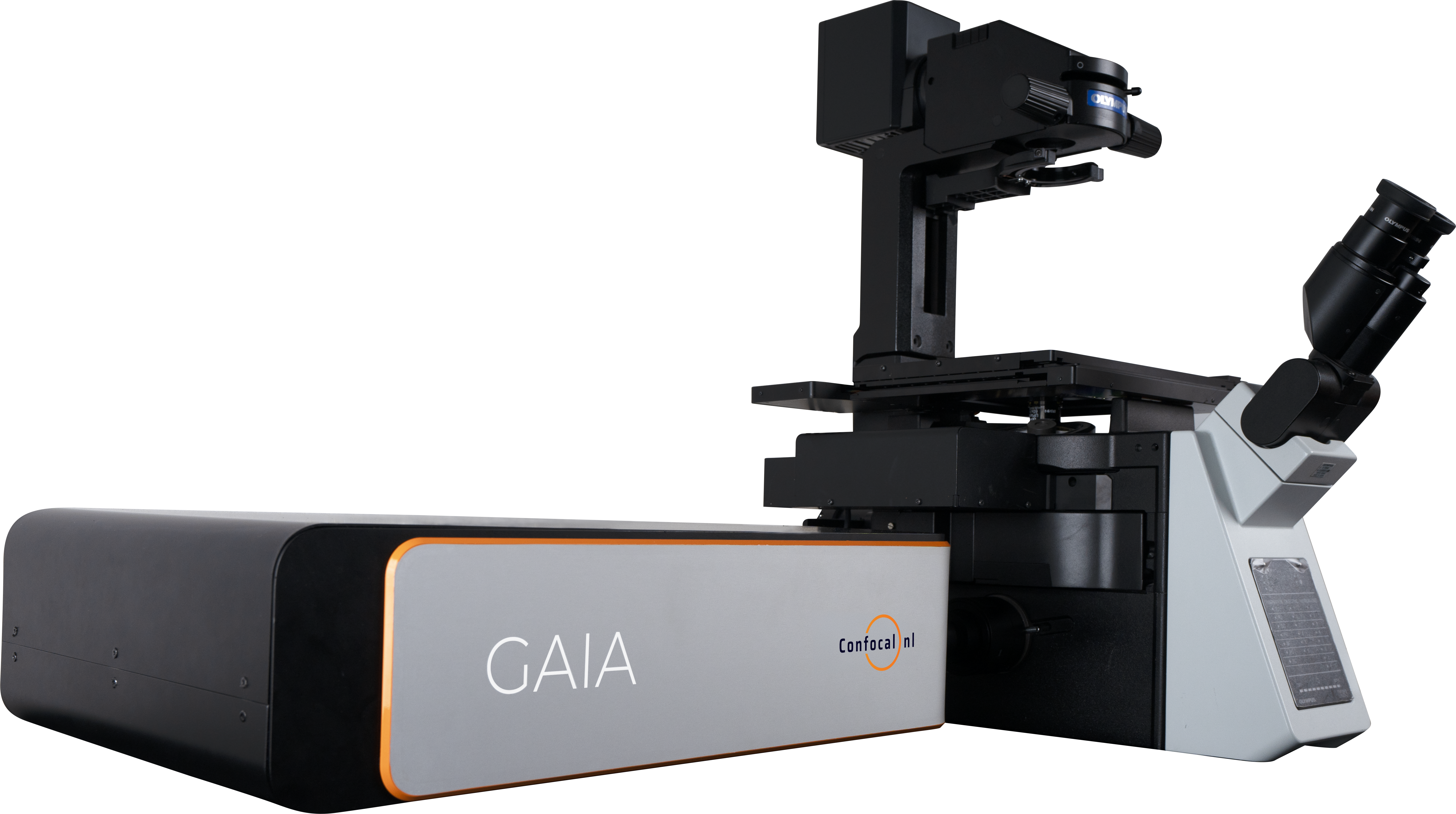
GAIA
The perfect solution for live cell super resolution imaging thanks to its very low phototoxicity.
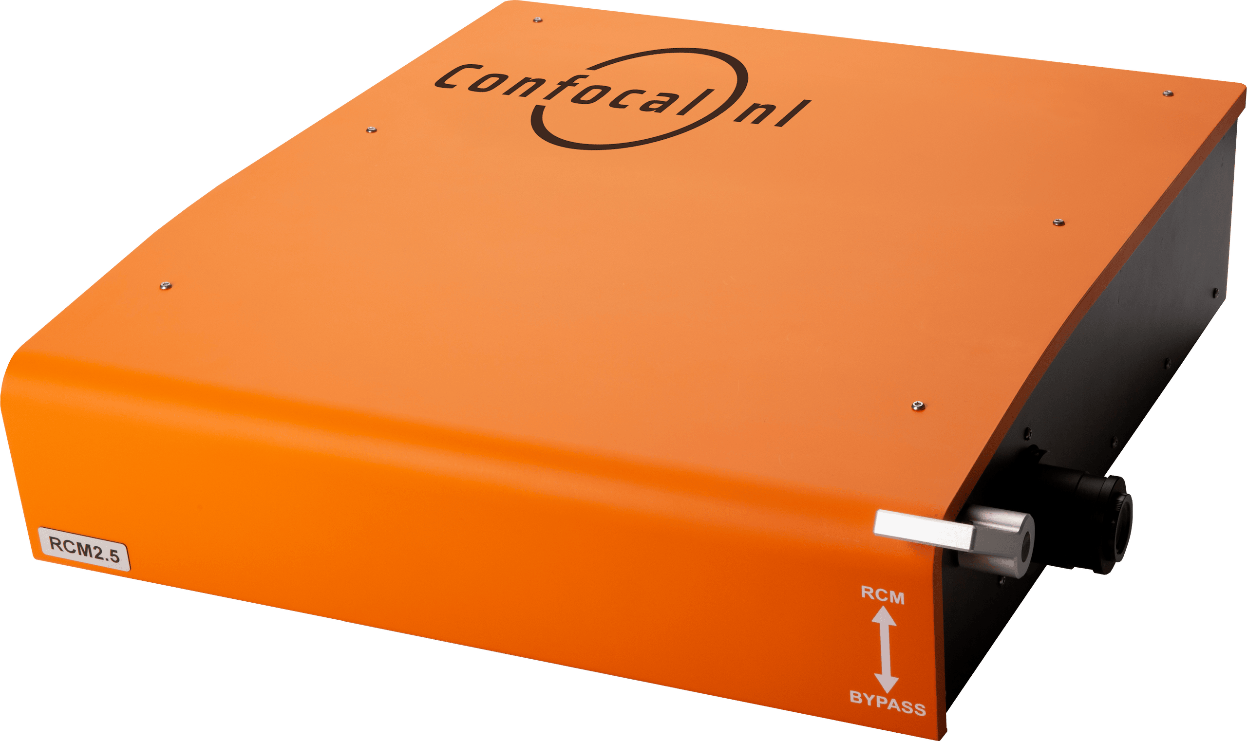
RCM
Capture datasets with 170nm raw resolution (120nm after deconvolution) by using 60x to 100x high numerical aperture (NA) magnification objectives and keep the laser intensity at a minimum.
NL5

NL5
NL5 is a fast confocal system with high sensitivity and resolution. Quickly screen a multi-well plate with multicolor images, and select the most promising ones.
AION

AION
AION is the third generation of our fast confocal technology. It provides high contrast images from thicker specimens such as organoids, tissue samples, plant and animal model organisms.
GAIA

GAIA
The perfect solution for live cell super resolution imaging thanks to its very low phototoxicity.
RCM
RCM
Capture datasets with 170nm raw resolution (120nm after deconvolution) by using 60x to 100x high numerical aperture (NA) magnification objectives and keep the laser intensity at a minimum.


