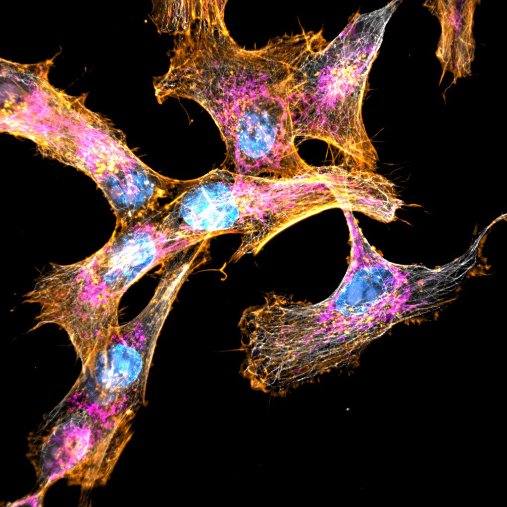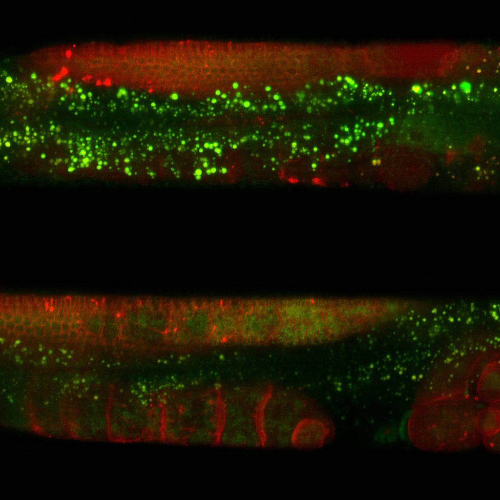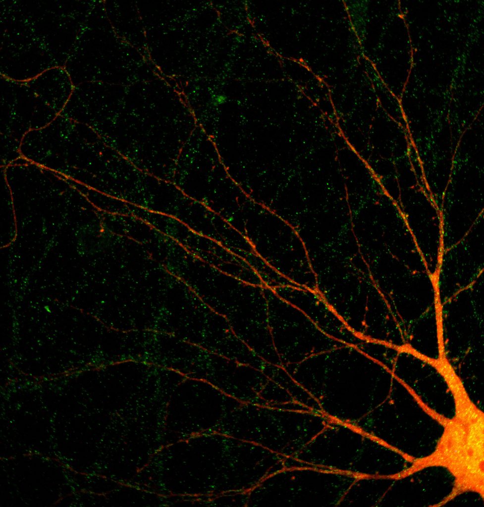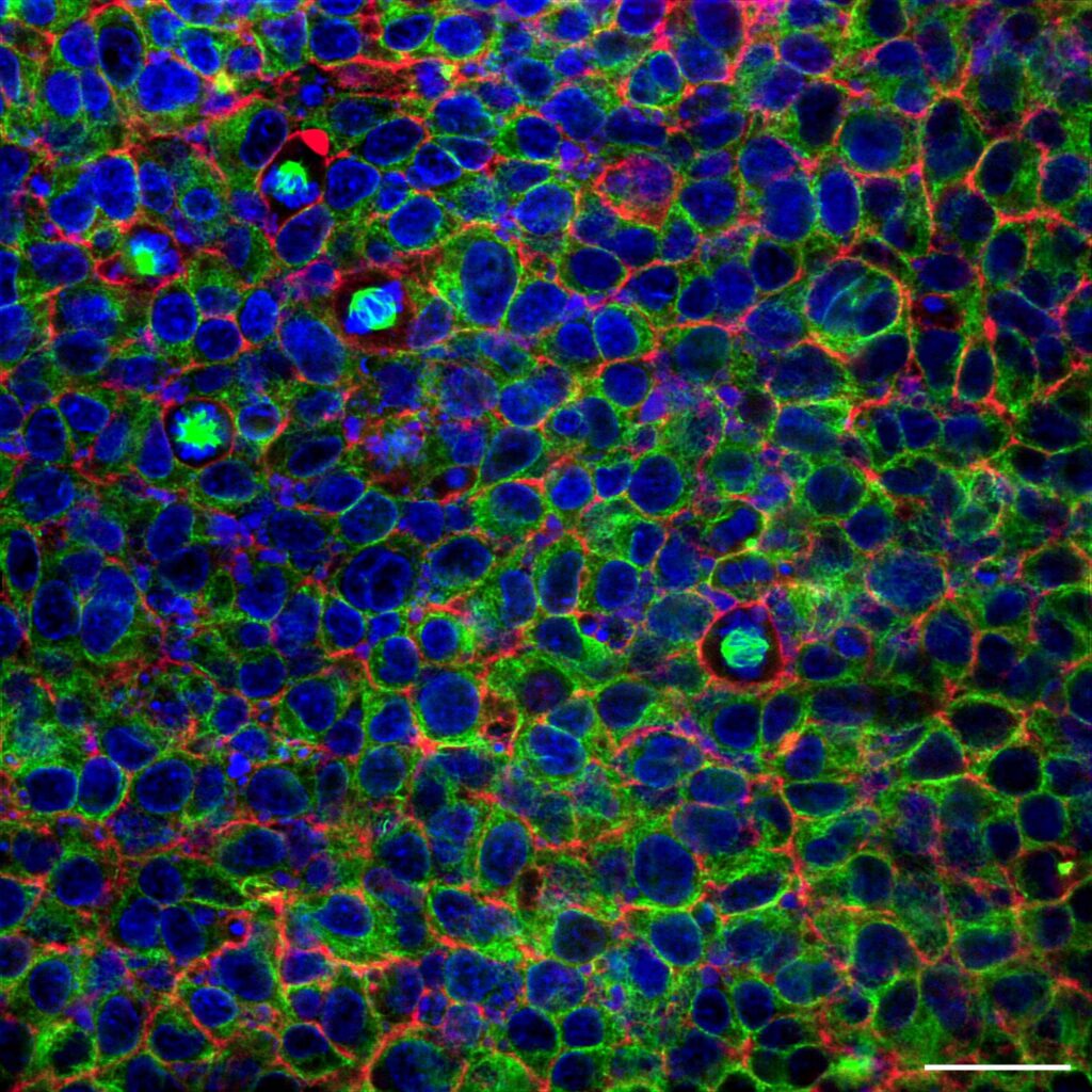


Tissue clearing provides researchers with a tool to investigate intact biological structures in three dimensions. This is achieved by making tissues transparent while preserving their cellular architecture and fluorescence. Tissue clearing removes lipids and other opaque elements, enabling deeper light penetration and clearer imaging. Combined with advanced microscopy techniques like multiphoton or light-sheet microscopy, cleared tissue imaging provides detailed views of complex systems, such as neural networks or vascular structures, within whole organs or large tissue sections. It is especially valuable in neuroscience, developmental biology, and pathology, where understanding spatial relationships and large-scale structures is crucial.
Cleared tissue
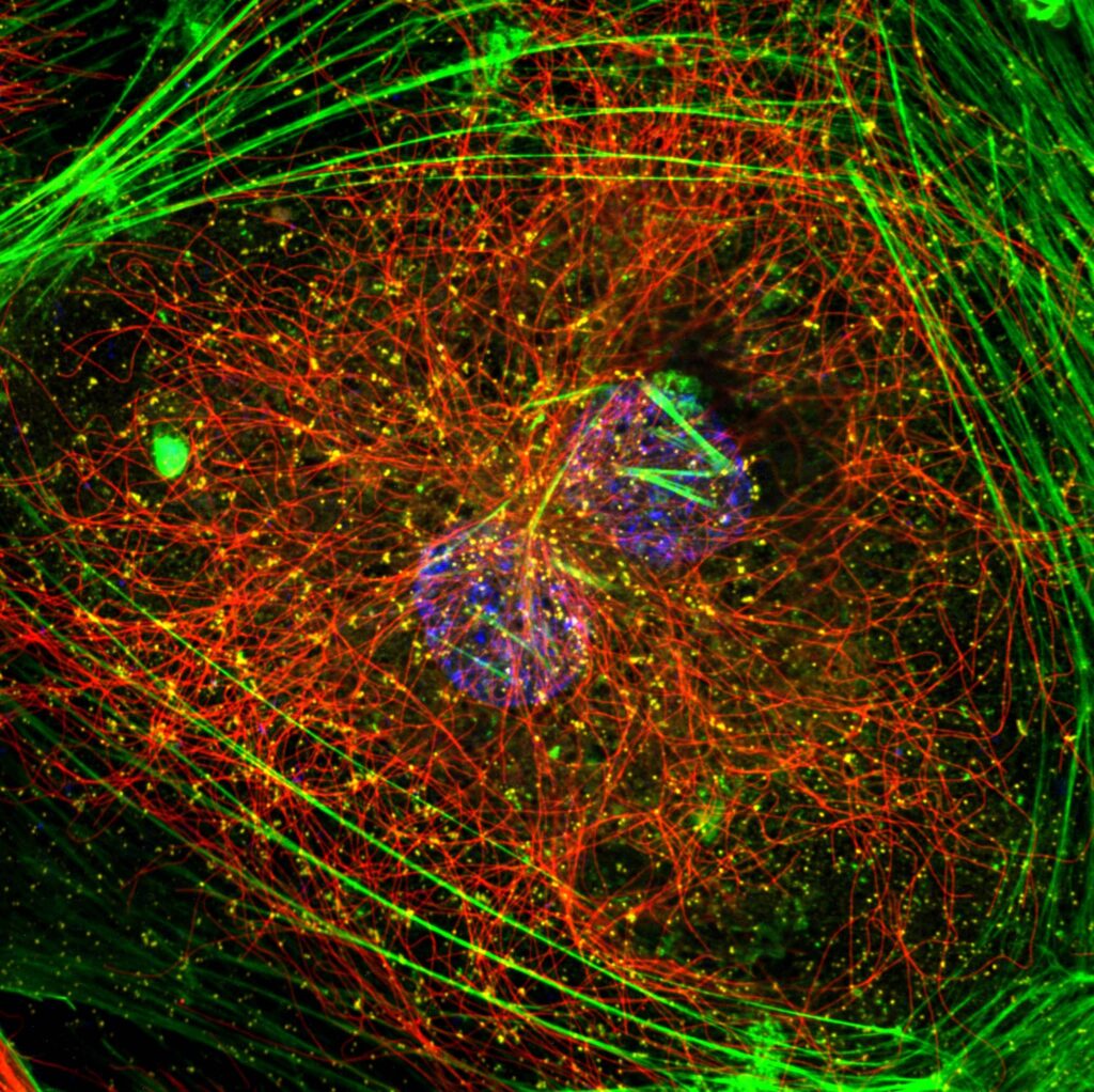
Super resolution microscopes that enable deep live cell imaging beyond the diffraction limit using only nanowatts of power.
Investigate intact biological structures
Unparalleled, in-depth view
Although clearing and imaging large tissues can be time-intensive and require specialized setups, it provides an unparalleled, in-depth view of biological organization. Confocal microscopy is not standardly used for cleared tissue imaging due to its limited depth penetration, as even cleared tissues can scatter light. The high laser power required for thick samples can cause photobleaching and photodamage. Additionally, the time-intensive scanning needed for large volumes may limit imaging speed and increase data processing demands.
An affordable alternative to multiphoton microscopy
NL5+ presents an affordable alternative to complex, expensive and delicate techniques like multiphoton and light-sheet microscopy. All the obstacles to using confocal microscopy for cleared tissue imaging, like limited depth, high laser power, and time-intensive scanning, can be overcome by using Line Rescan NL5+. The only criterion that needs to be met is the adequate positioning of the sample.
Products
Discover our solutions
Find the appropriate confocal system for your imaging needs.
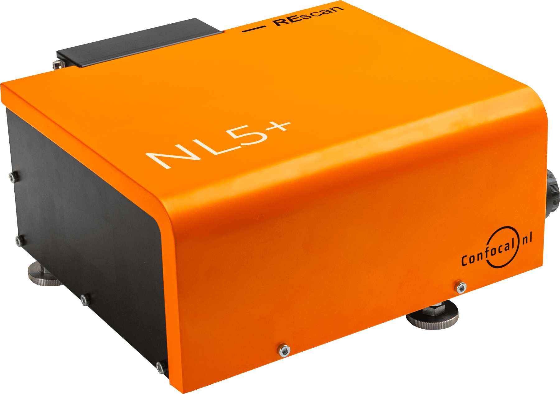
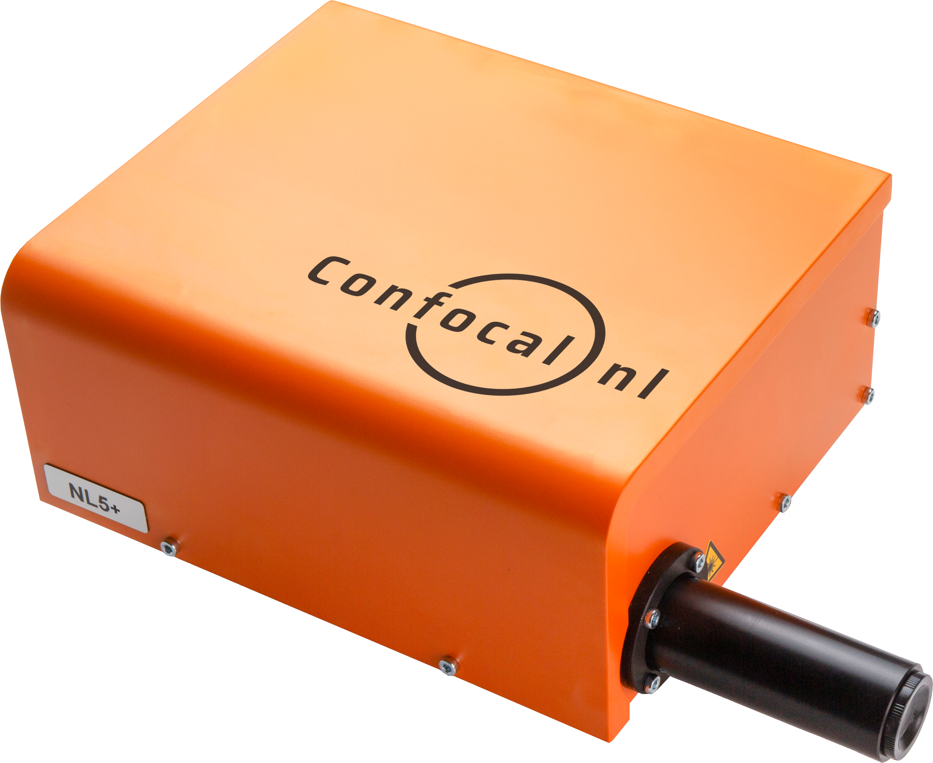
NL5+
NL5+ is a fast confocal system with high sensitivity and resolution. Quickly screen a multi-well plate with multicolor images, and select the most promising ones.
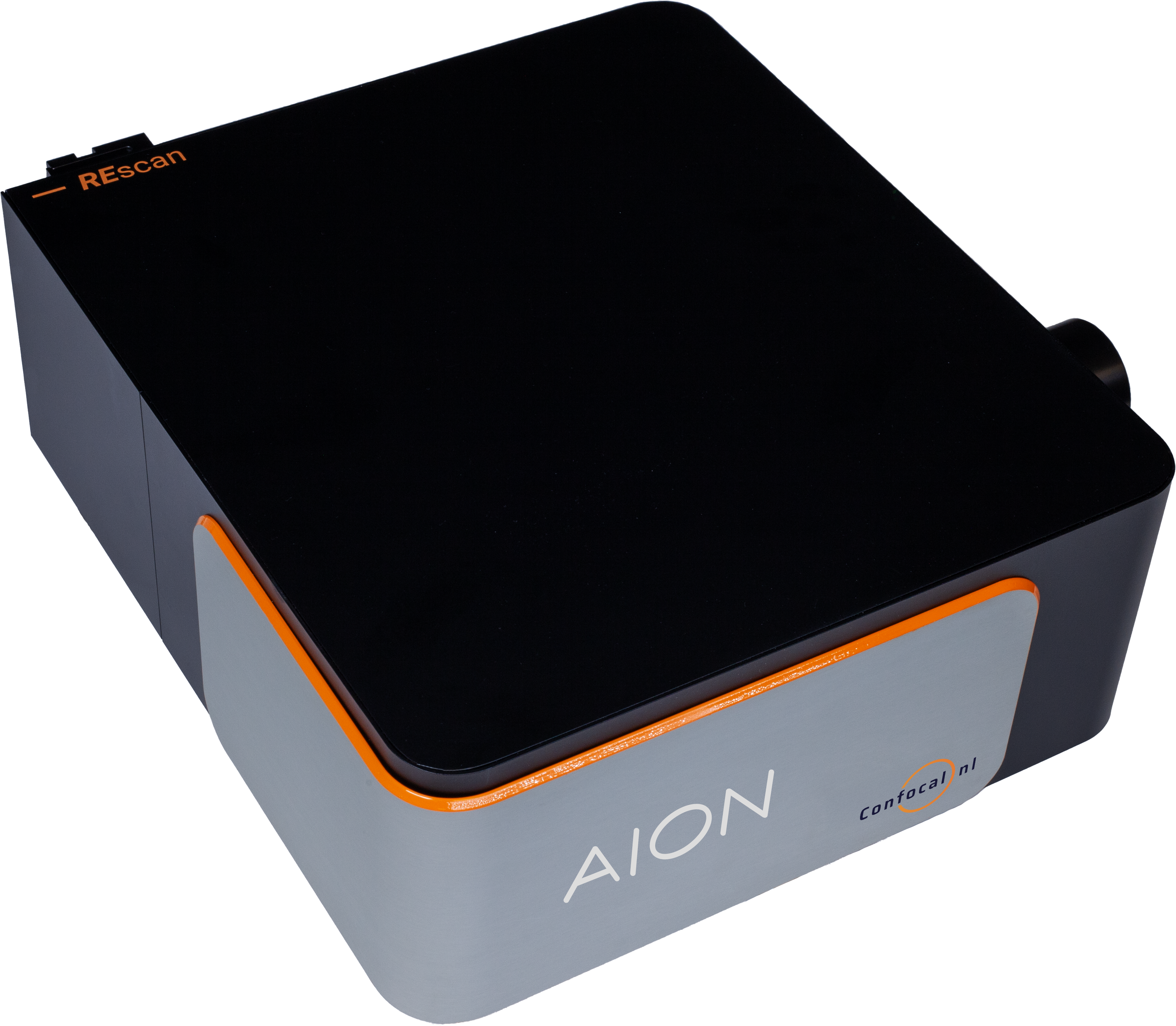
AION
AION is the third generation of our fast confocal technology. It provides high contrast images from thicker specimens such as organoids, tissue samples, plant and animal model organisms.
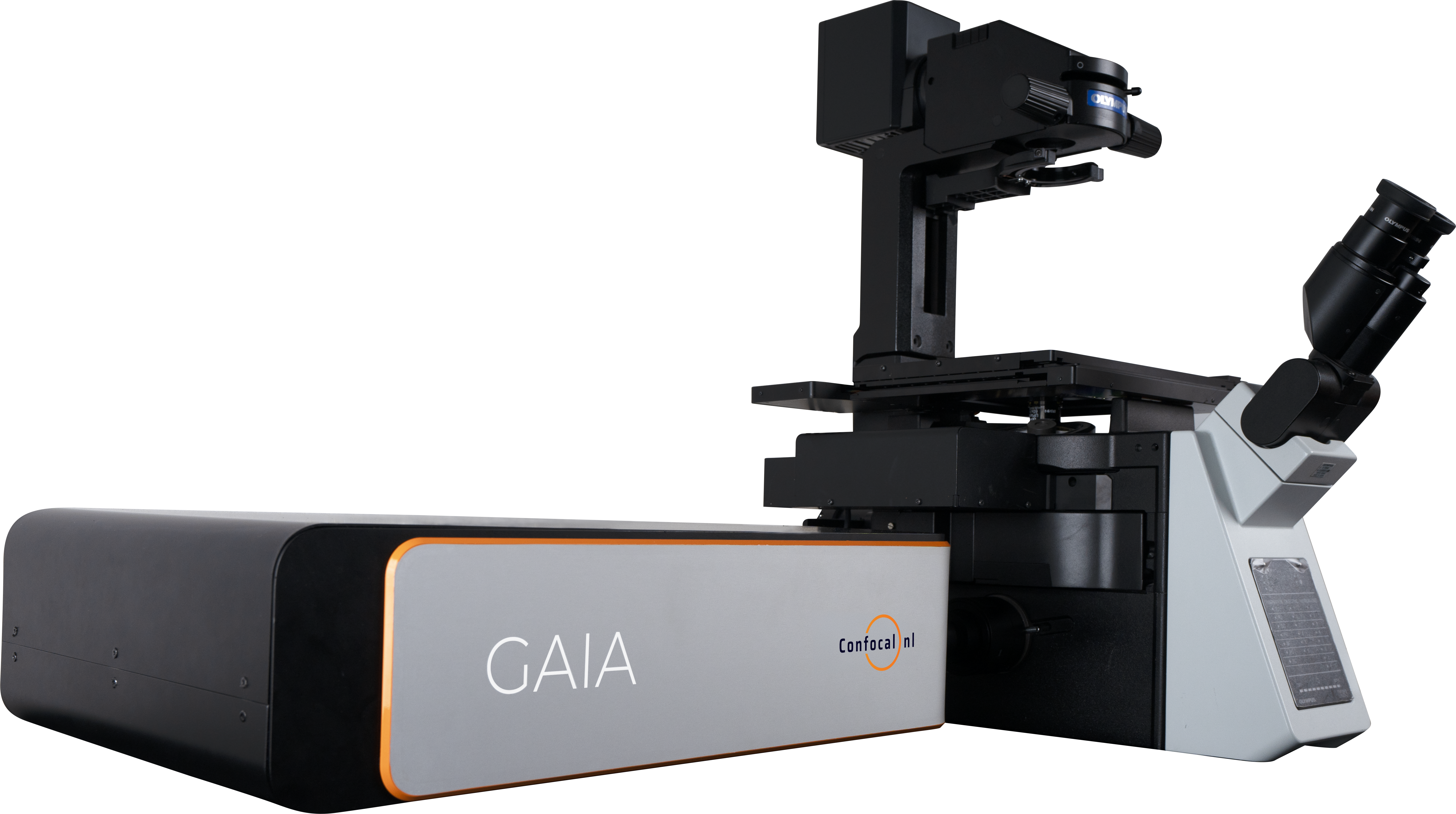
GAIA
The perfect solution for live cell super resolution imaging thanks to its very low phototoxicity.
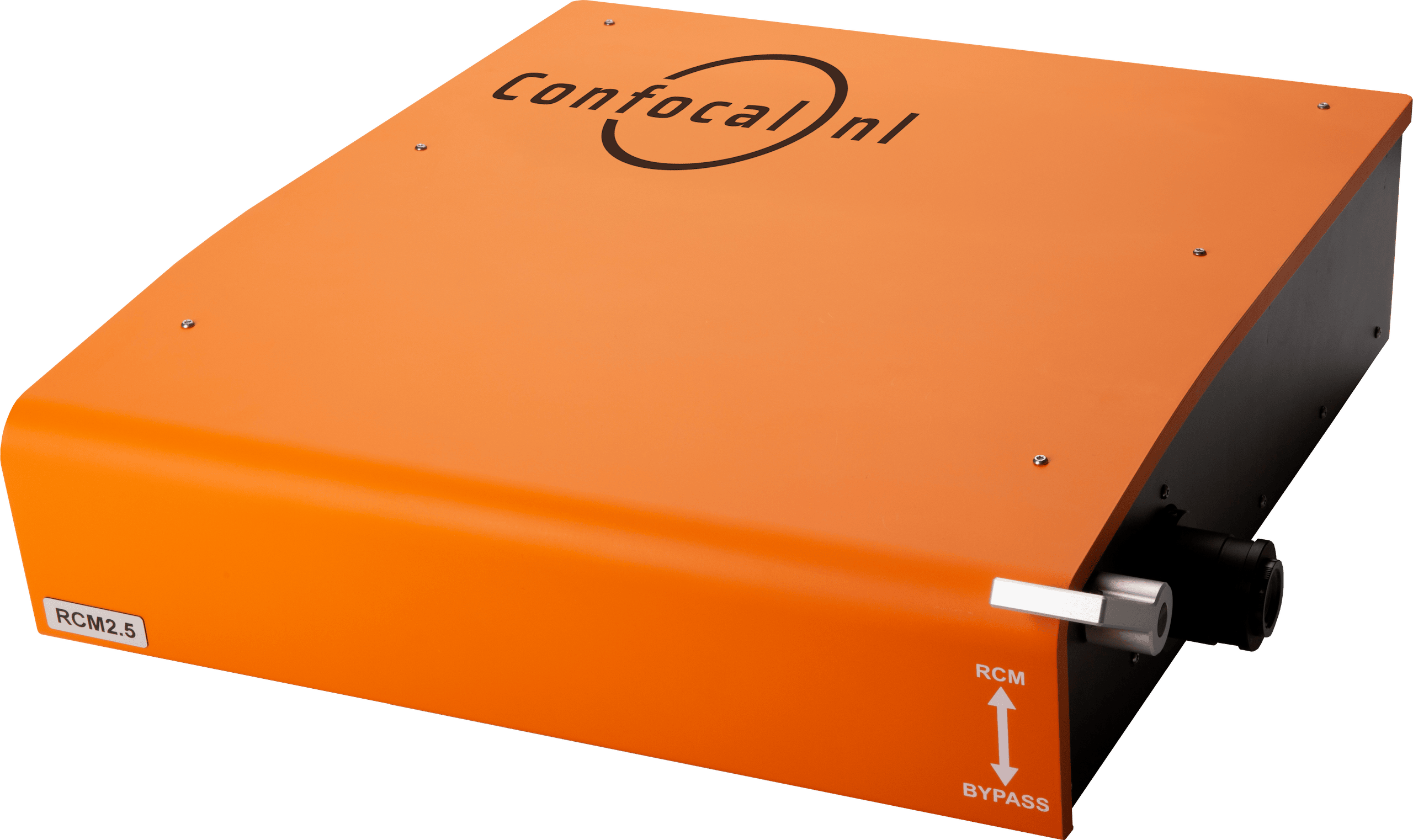
RCM
Capture datasets with 170nm raw resolution (120nm after deconvolution) by using 60x to 100x high numerical aperture (NA) magnification objectives and keep the laser intensity at a minimum.
KRONOS
KRONOS enables researchers to maximise the output of their REscan confocal system. It unlocks the system’s full potential for an improved imaging experience.
ZEN Module
Operate your AION and NL5+ Line REscan confocal microscopes directly within the familiar ZEISS ZEN environment and bring high speed confocal imaging into the workflow researchers already trust.
NL5
NL5
NL5 is a fast confocal system with high sensitivity and resolution. Quickly screen a multi-well plate with multicolor images, and select the most promising ones.
AION
AION
AION is the third generation of our fast confocal technology. It provides high contrast images from thicker specimens such as organoids, tissue samples, plant and animal model organisms.
GAIA
GAIA
The perfect solution for live cell super resolution imaging thanks to its very low phototoxicity.
RCM
RCM
Capture datasets with 170nm raw resolution (120nm after deconvolution) by using 60x to 100x high numerical aperture (NA) magnification objectives and keep the laser intensity at a minimum.
Resources