



“Some neurons of the tiny worm C. elegans carry cilia: small antennas that let the animal ‘taste’ chemicals in its environment.” Prof. Erwin Peterman (Vrije Universiteit Amsterdam) and his team study how these cilia work in living worms, from single molecules all the way to the animal’s behaviour.
In this video they explain:
- How proteins reach the right place in the cilium through a mix of motor-driven intraflagellar transport and random diffusion.
- How a single molecule binding to a receptor can trigger neuronal activity and change behaviour.
- Why this research needs fluorescence microscopy that is sensitive, specific, fast and above all gentle, to avoid photobleaching and phototoxicity.
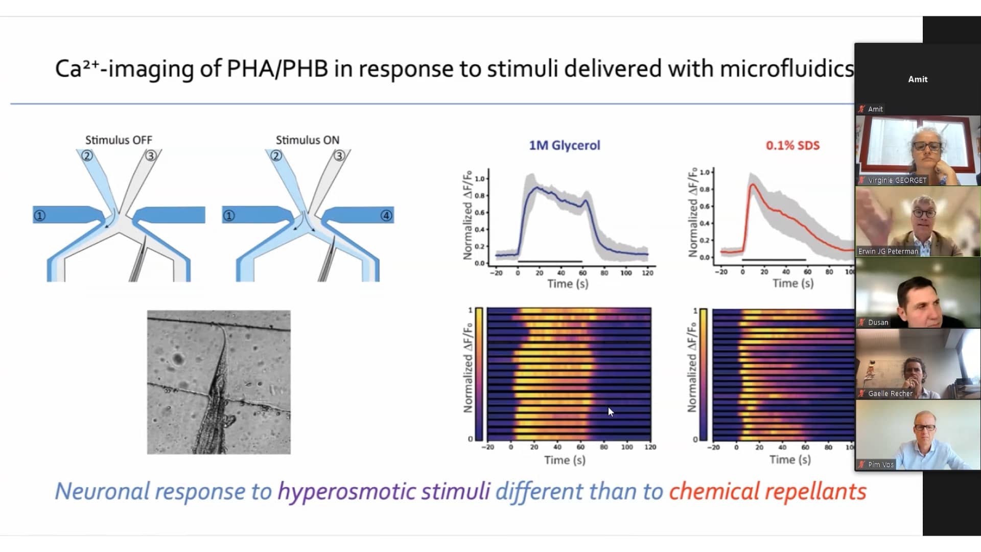
- How worms in a microfluidic chip are exposed to chemicals while the response of the cilia and nervous system is imaged.
The team describes how the NL5+ Line REscan confocal lets them image thicker parts of the worm with less background, record very long image sequences, and adjust settings easily.