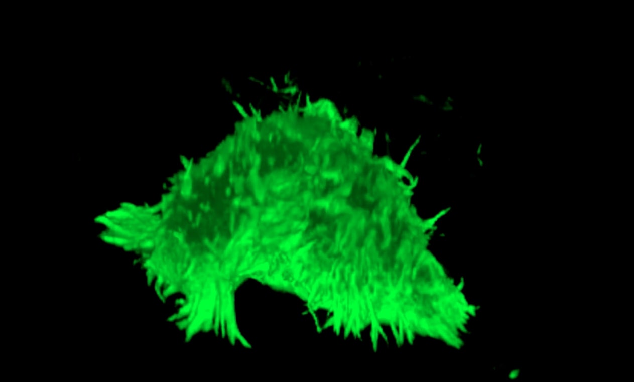
Life cell imaging is the way to go when you want to get insights into dynamic processes occurring inside and between cells. With the help of fluorescent labels, molecule localization and cell-to-cell interactions can be recorded in real-time. One of the main obstacles during life cell imaging is the damage caused by light (phototoxicity or photobleaching). This can happen due to the use of high-intensity light or by prolonged exposure to it. Light of low or high wavelength can damage cells by increasing temperatures or the generation of reactive oxygen species (ROS). Fluorophores used to visualize structures/molecules inside the cell produce ROS when excited by light. This can directly damage proteins, affect cell morphology or even lead to cell death. This process
is called phototoxicity.
In this blog, we’ll tell you more about the art of cell imaging and how to find the balance between cell survival and image quality!
Would you like to know more about phototoxicity? Dr. Erik Manders will tell you all about it in our latest webinar: ‘How to prevent phototoxicity.
How have our products won the race against phototoxicity?
Researchers often use photomultiplier tubes (PMT) as light detectors in conventional confocal microscopy. PMTs capture incident photons and generate electronic charges that are amplified. The relative efficiency of light detection from a detector is often expressed as quantum efficiency (QE) as a function of wavelength. As you will see in the graph below, the efficiency of this process is not optimal. Available PMTs in the market have a relative efficiency ranging from 40 % QE at the visible range of light to 20% at the Near-Infrared wavelength.

To avoid this loss of sensitivity, Confocal.nl has replaced PMTs detectors for digital cameras using sCMOS sensors or other camera techniques like EMCCD. The quantum efficiency (QE) has then been increased by up to 95% in the visible range of light and to 67% in the Near-Infrared. This increase in sensitivity allows live cell imaging with very low laser intensities. Which then results in less
phototoxicity and bleaching.
Replacing PMTs with high sensitivity cameras like sCMOS or EMCCD allows us to use less laser power to obtain high-resolution images in real-time.
Discover more about RCM!
RCM is excellent for biological applications where super-resolution and high sensitivity are required. The clever design of RCM makes these products robust, versatile, and extremely easy to use. Do you want to know how RCM can improve your imaging experience? There is enough to learn!


