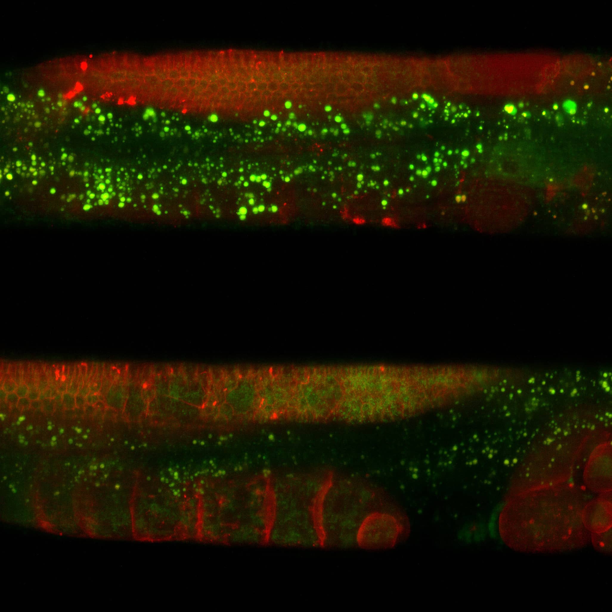
The larval zebrafish serves as a powerful model for studying neural circuit development due to its transparency and suitability for fluorescent microscopy. Using GAIA Point REscan confocal microscope, we performed super resolution imaging to investigate the structural and functional development of the zebrafish nervous system between three and five days post-fertilization. GAIA Point REscan facilitated extended imaging sessions with minimal phototoxicity, offering new insights into vertebrate neural development. Imaging revealed key features, such as synaptic boutons and axonal projections, critical for circuit formation. Combining super resolution structural imaging with functional assays enabled the dynamic tracking of neural rewiring.
Download the application note
Learn more about this application.


