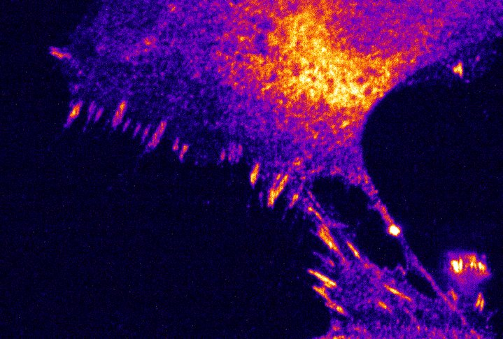
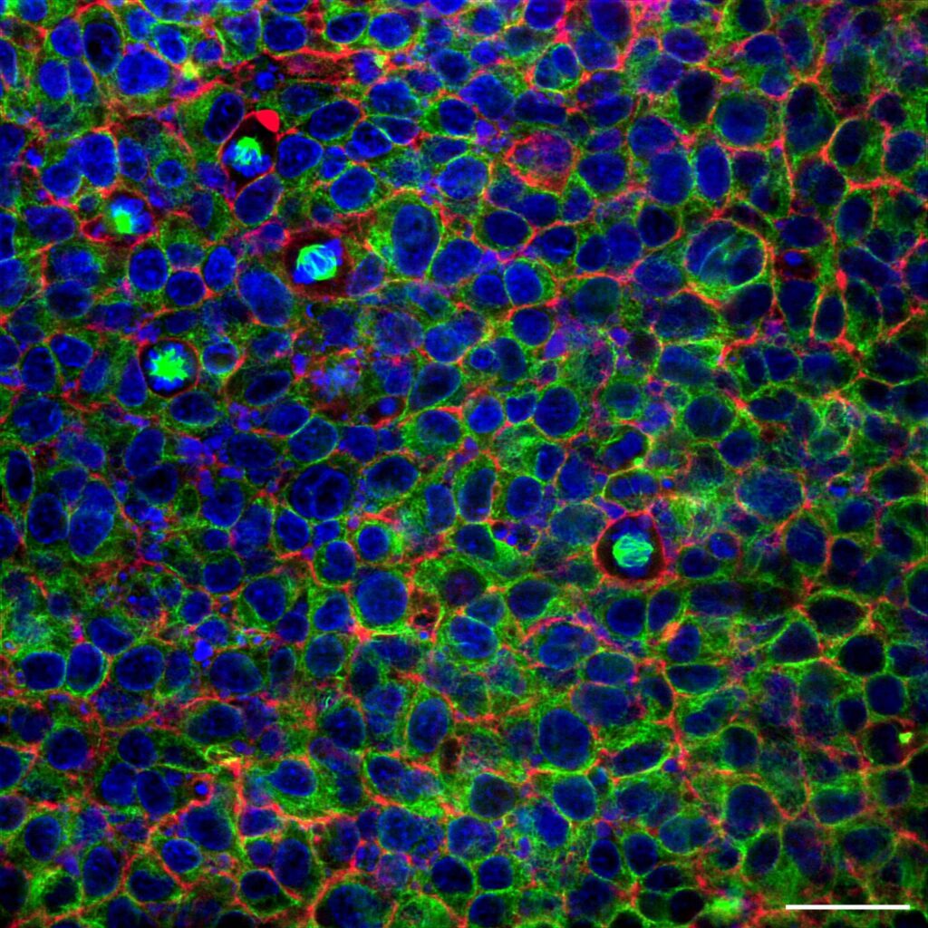
KRONOS

Turning complexity into clarity

REscan imaging – easier than ever with KRONOS

Confocal microscopy keeps opening new frontiers of discovery, but it can be complex to operate. We developed our microscopy acquisition software to change that. With an intuitive interface, streamlined workflows, and powerful imaging capabilities, KRONOS removes the friction from acquisition so scientists can focus on what matters most: their research.

KRONOS enables researchers to maximise the output of their REscan confocal system. It unlocks the system’s full potential for an improved imaging experience.

Request a demo

Powerful imaging effortlessly

Full control of your REscan confocal microscope
You can easily control your REscan confocal unit, lasers, cameras, and the microscope body, with seamless switching between confocal, bypass, and widefield modalities.

Self-adjusted laser power
Regardless of the imaging depth,  KRONOS can self-adjust your laser power to ensure constant gentle imaging without compromising signal-to-noise.

Live feedback from your experiment
Get real-time image preview with adjustable parameters, allowing you to optimize acquisition instantly.

Seamless data export
Open your acquisition data set in FIJI instantly—just one click to transfer and explore.

Multidimensional imaging
Easily navigate across x, y, z, time, and lambda dimensions with multiposition stage control, perform tile scans or custom regions within the field of view.

Intuitive interface

Master in 5 mins

Power in depth

Self-adjust laser power

GPU Powered

Render images in real time

Seamless stitching

Top-class uniformity

Set up and image in minutes

Easy to Learn, Easy to Use
Become a pro in five minutes and set up your experiment. Designed with scientists in mind.

Future-Proof
Flexible to support emerging imaging techniques and applications. Designed to accept future compatibilities.

Research Accelerated
From acquisition to analysis, KRONOS helps researchers work smarter and faster.

Trusted Performance
Built on reliable, proven imaging standards.

Applications

Cell biology
Image cells and their components in detail—from organelles to proteins—using confocal and super-resolution microscopy, driving discoveries in cell biology and drug development.

Developmental biology
Track cell differentiation and organ development in real time, and study signalling pathways.
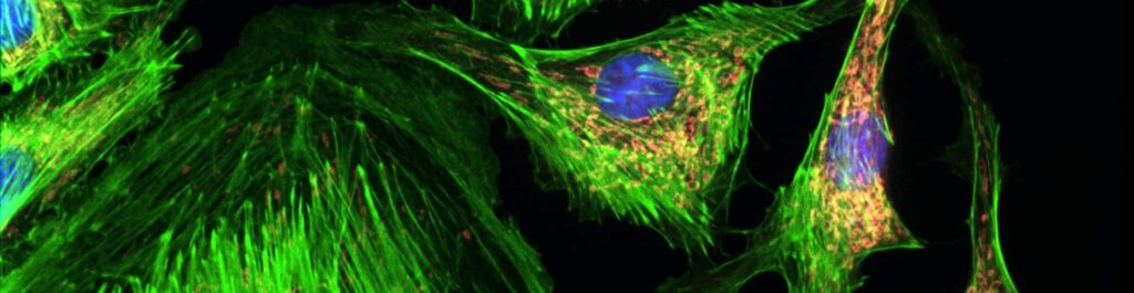
Live cell imaging
Study living cells in real time with fluorescence, confocal, and time-lapse microscopy to visualize dynamic processes like division, migration, trafficking, and signalling while preserving cell health.

Deep Live cell imaging in Super Resolution
Image beyond the diffraction limit with super-resolution microscopy, revealing nanoscale structures, protein interactions, and dynamic processes that transform our understanding of health and disease.

Neurobiology
Capture synaptic connections in fine detail with confocal and super-resolution microscopy, revealing nanoscale structures and protein localisation to advance understanding of brain function and disease.

Plant Biology
Visualize complex three-dimensional plant structures with confocal microscopy, from root tips to leaf tissues, and track dynamic processes like photosynthesis, nutrient transport, and cell division.

Ca²⁺ imaging
Monitor real-time changes in intracellular calcium concentrations with calcium imaging, revealing critical signalling processes in neuroscience and cardiac research.

C. elegans
Study key biological phenomena of C. elegans by imaging its transparent body and cellular structures in real time, from development and behaviour to neural networks, gene expression, and nanoscale synaptic details.
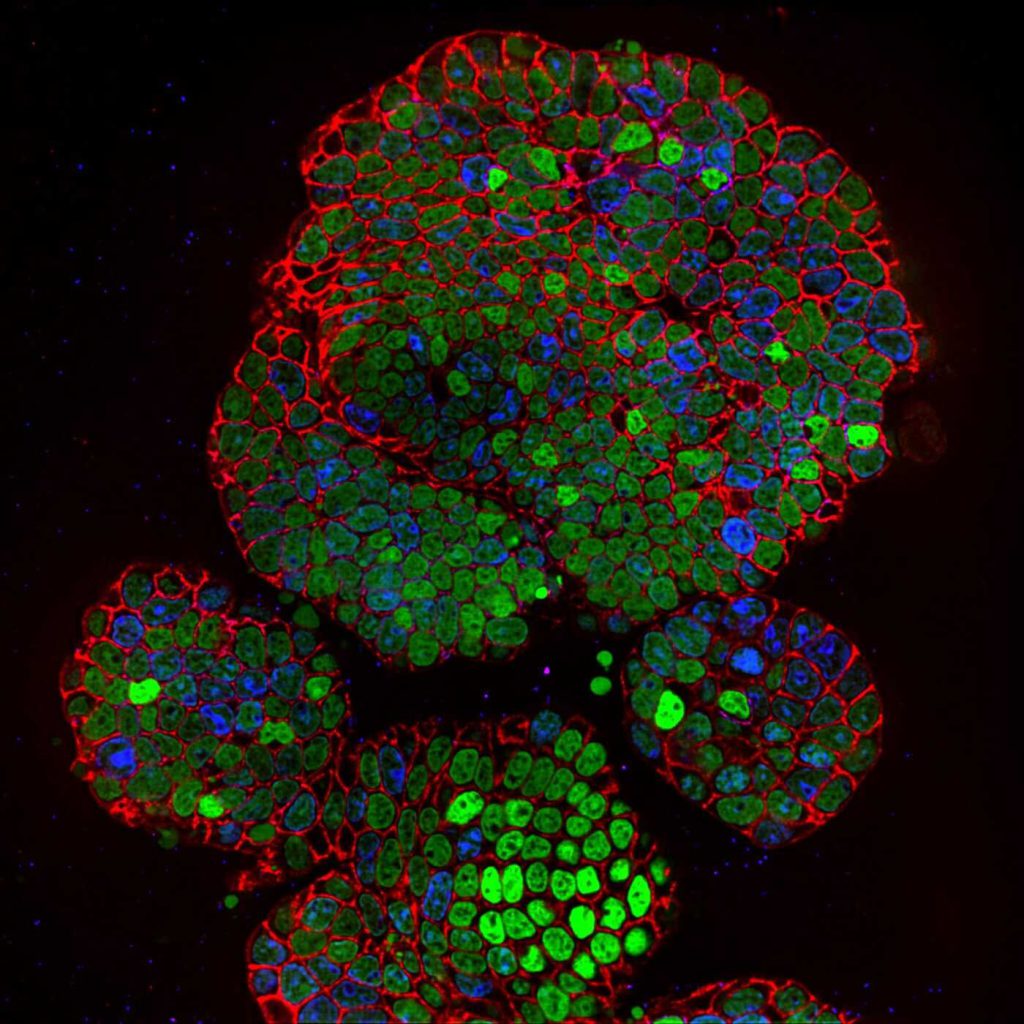
Organoids
Observe cellular interactions in a realistic environment by imaging organoids, enabling studies of development, disease, drug testing, and personalized medicine in models that closely mimic human biology.
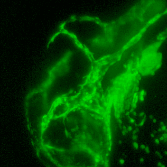
Cleared tissue
Investigate intact biological structures in three dimensions with tissue clearing, preserving cellular architecture while enabling deep imaging of complex systems like neural networks and vascular structures.

Compatibility & recommended hardware

Micrsocope Body Compatibility
KRONOS is compatible with most commercially available microscope bodies. Contact us for specific configurations or for the full compatibility list. 

Processor
Intel Core i7-13700 processor or higher.

Memory
64GB RAM or higher. 

Graphics Card
NVIDIA GeForce RTX 4060 or Quadro A4000. 

Storage
1TB or more NVMe SSD with a separate mechanical backup hard drive to store backups of KRONOS’s configuration files.

Operating System
Windows 11. 

Display
A minimum of 2560×1440 resolution and IPS panel technology for optimal image viewing.

Testimonials

Satisfied researchers
around the globe

Prof. Hei Ming LaiChinese University of Hong Kong

The NL5 allowed us to image 500 μm deep into cleared brain tissue samples with a high-signal-to-noise ratio, something that was difficult to achieve with other techniques.
This, in combination with its gentle imaging capabilities, high acquisition speed, and intuitive interface, made it possible to easily observe our samples.

Prof. Erwin PetermanFaculty of Science, Physics of Living Systems, Vrije Universiteit Amsterdam, The Netherlands

The NL5 has enabled us to image deep in different parts of the C.elegans that we could not so far measure properly.

Gabor CsucsETH Zürich

RCM is a very easy to use system that we have in combination with STORM and it provides great underlay images for STORM data.

Dr. Yannick LeursChemical Biology, Eindhoven University of Technology

The NL5+ has been instrumental in advancing our studies. It allowed us to image our samples quickly and deeply with very low laser power, providing consistent, high-quality imaging.

Resources

Related Resources

Advantages of Re-scan confocal microscopy
Confocal blog
Advantages of Re-scan confocal microscopy

Advantages of Re-scan confocal microscopy

RCM high contrast images allow for optimal deconvolution. Find out more, download the full document.
Downloadable documentNewsMay 4, 2021 Read More
Advantages of Re-scan confocal microscopy
Confocal blog
Advantages of Re-scan confocal microscopy

Advantages of Re-scan confocal microscopy

RCM high contrast images allow for optimal deconvolution. Find out more, download the full document.
Downloadable documentNewsMay 4, 2021 Read More

Download the product flyer

Receive more information on this product or request a quote.

Request a quote