




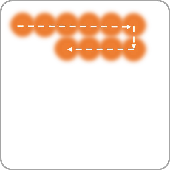
REscan comes in two types: Point REscan (RCM2.5, RCM2, RCM1) and Line REscan (NL5+, NL5). In this second Quick Knowledge, we compare how each type illuminates the sample and what that means for your imaging. Point REscan breaks the diffraction limit for live cell super resolution (120 nm), while Line REscan images live cells at up to 75 fps. We also show why both need less illumination, which keeps phototoxicity low and lets you image up to 650 µm deep.
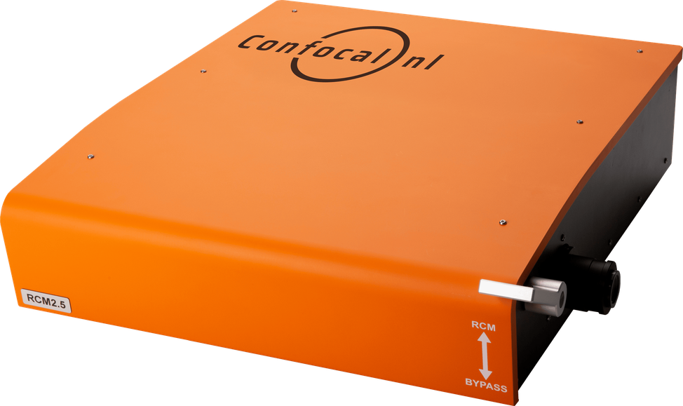
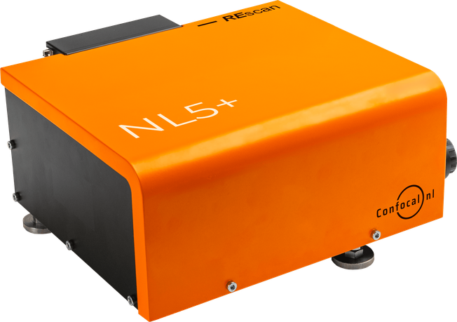
There are 2 types of
REscan Confocal units
Point REscan
Line REscan
RCM2.5
RCM2
RCM1
NL5+
NL5
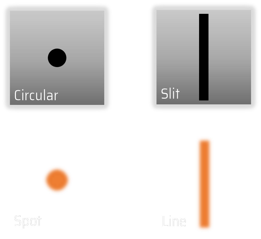
The difference
between REscans
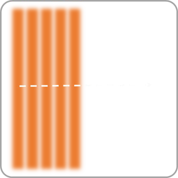
Point REscan
Line REscan
Pinhole
Illumination
Different REscans,
different benefits
Point REscan
Line REscan
Breaking the
diffraction limit
Live cell super
resolution (120nm)
Diffraction limited at
high speeds
Live cell imaging
up to 75fps
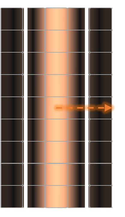
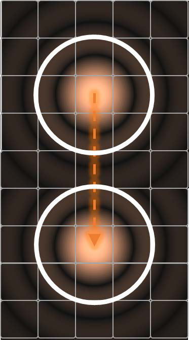
Is resolution
the same in X and Y
when using a slit?
Line REscan
Point REscan
The slit is basically a
small pinhole along
one axis and a wide
open pinhole along the
other axis.
In all REscans, pinhole
size has no influence on
the XY resolution,
because the detector is
pixelated (see Part 1).
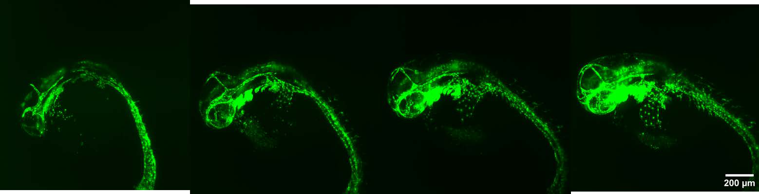
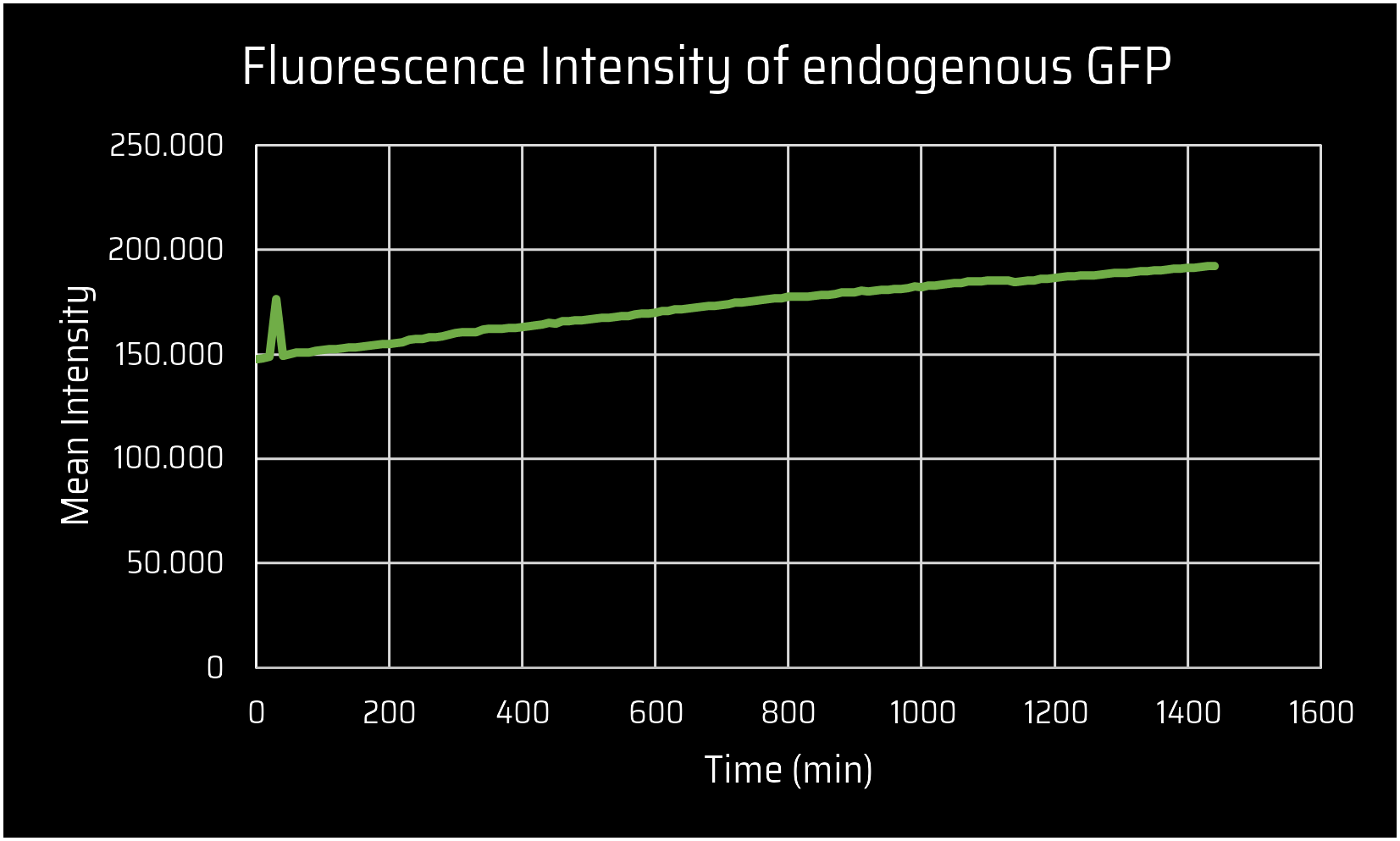
Low Phototoxicity of
REscan Confocal
Compared to standard confocals…
0 h
8 h
16 h
24 h
Growing zebrafish
REscan needs
way less
illumination...
making REscan
more cell
friendly!
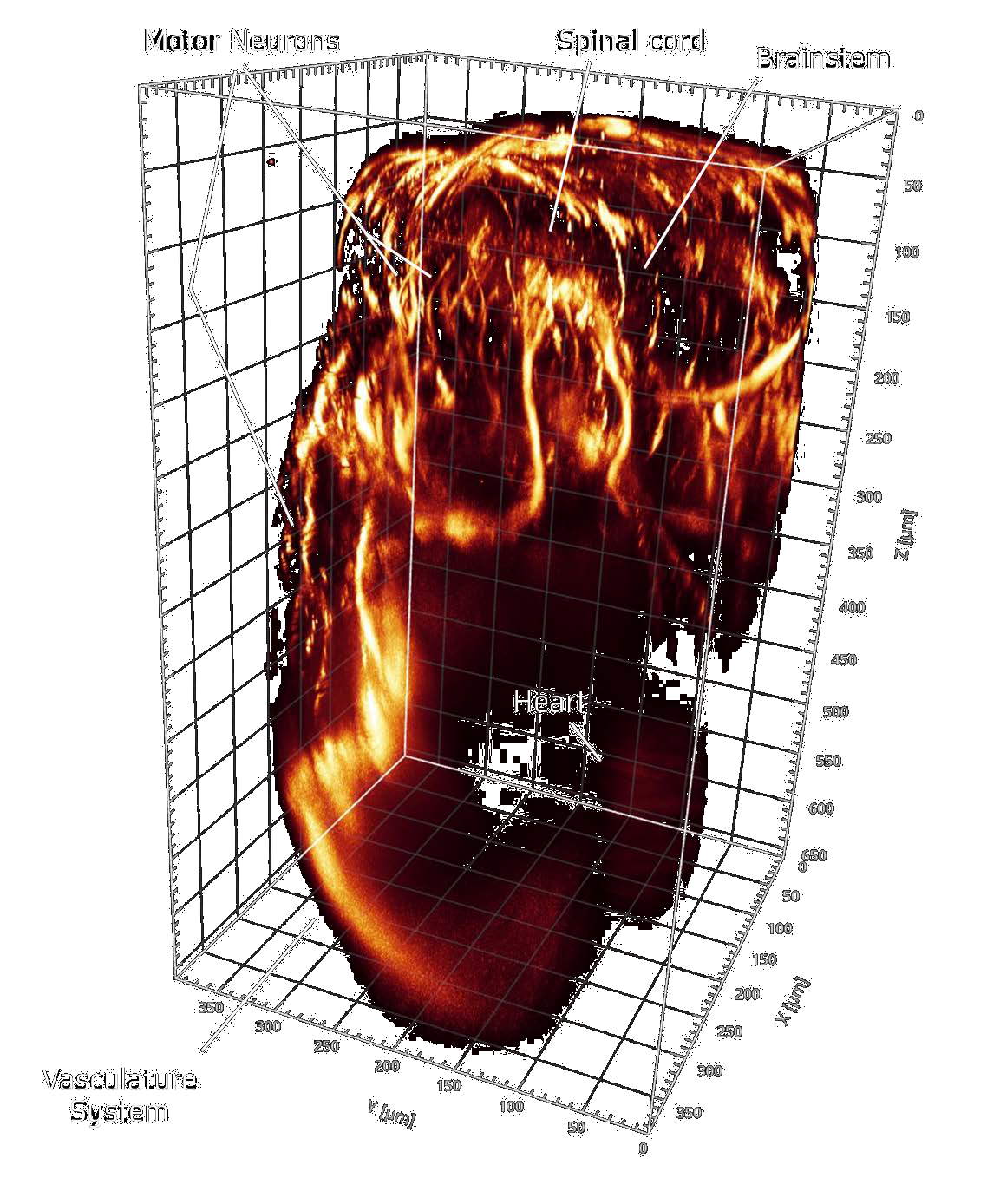
Deep imaging with
REscan Confocal
Less illumination
means less
bleaching of out
of focus cells
when going up to
650 µm in depth.
Zebrafish
Want to find out how REscan compares to
other confocal systems, especially in deep and
live cell imaging? Check Quick Knowledge Part 3!
Download the quick knowledge
Receive more information about this knowledge article.