


R M P Breedijk, J Wen, V Krishnaswami, T Bernas, E M M Manders, P Setlow, N O E Vischer, S Brul | Sci Rep. 2020 Mar 24;10(1):5312. doi: 10.1038/s41598-020-62377-1.
Summary
Time-lapse super-resolution imaging of living cells remains challenging, especially when photon signal is limited. Many current methods rely on specialized probes, high illumination, repeated image capture, or computational reconstruction.
In this study, the authors introduced an optics-based approach combining annular illumination with re-scan confocal microscopy. Because images are generated in a single scan, the method avoids reconstruction artifacts associated with multi-frame techniques.
The system achieved lateral resolution comparable to linear structured illumination microscopy, while maintaining axial resolution similar to a conventional confocal microscope.
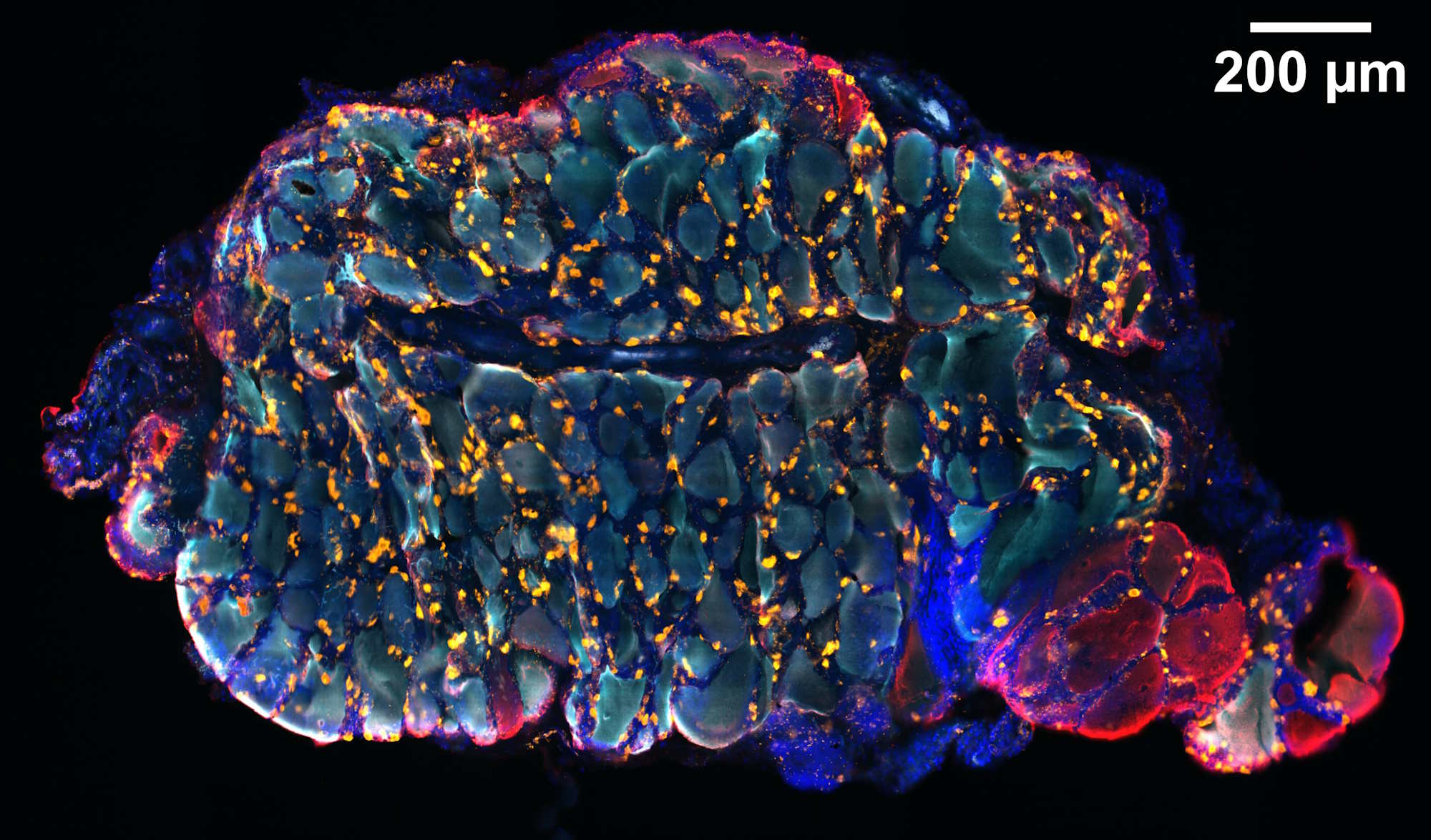
As a biological demonstration, the authors applied the method to wild-type Bacillus subtilis spores containing germination receptor clusters known as germinosomes. They provided the first evidence of germinosomes in wild-type spores, revealed their spatial and temporal dynamics after germinant exposure, and visualized the early stages of spore revival.
Read the publication: https://pubmed.ncbi.nlm.nih.gov/32210351