


Gambarotto, D., Zwettler, F. U., Le Guennec, M., Schmidt-Cernohorska, M., Fortun, D., Borgers, S., Heine, J., Schloetel, J. G., Reuss, M., Unser, M., Boyden, E. S., Sauer, M., Hamel, V., & Guichard, P. (2018) | Nature Methods, 16(1), 71–74
Summary
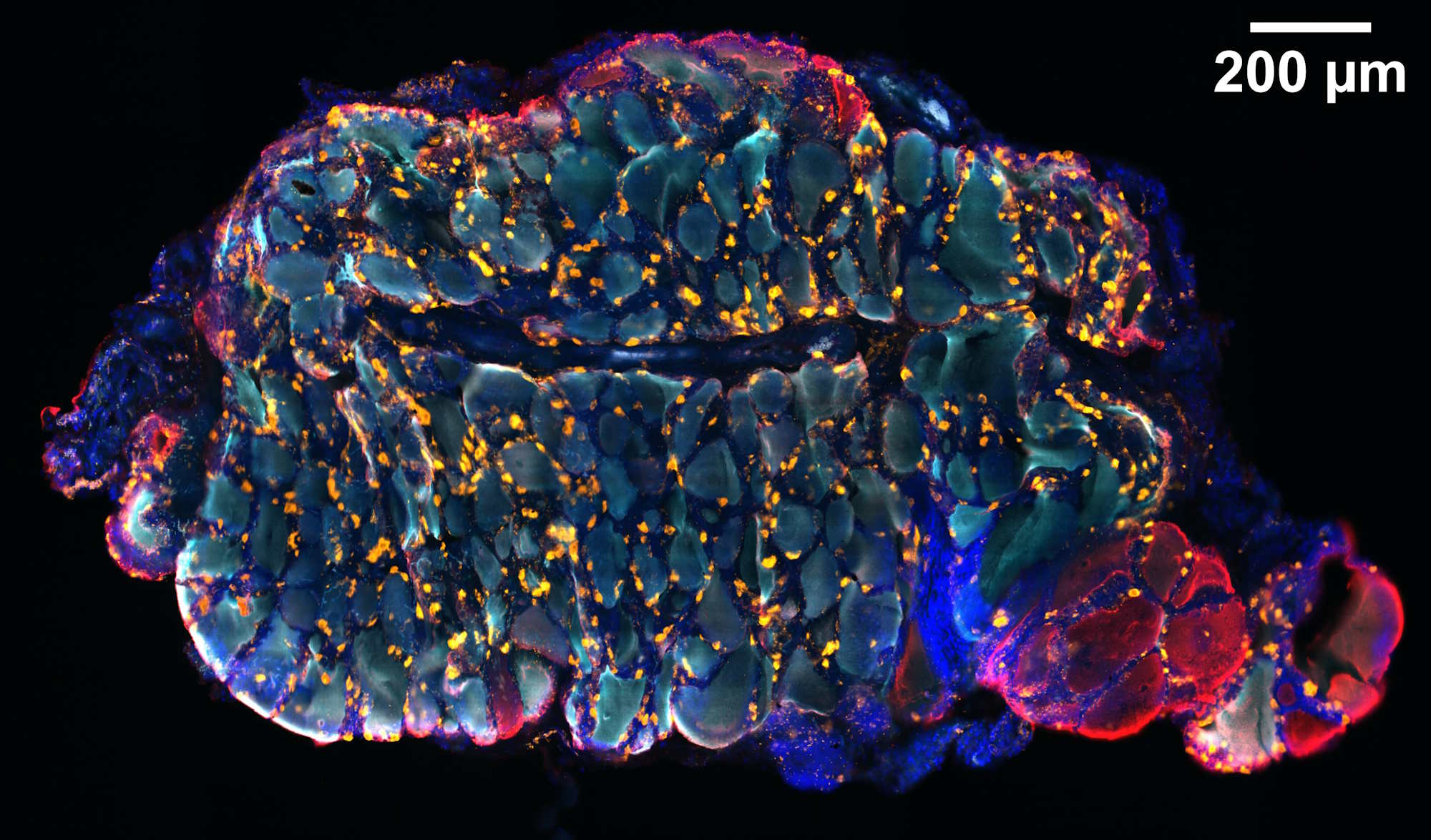
Determining the structure and composition of macromolecular assemblies remains a major challenge in biology. In this study, the authors introduced ultrastructure expansion microscopy (U-ExM), an advanced form of expansion microscopy designed for optical imaging of preserved ultrastructural features.
The method enables near-native expansion of biological structures both in vitro and within cells, making fine details accessible through conventional optical microscopy. When combined with super-resolution imaging, U-ExM revealed cellular ultrastructural features such as centriolar chirality that were previously visible only by electron microscopy.
The findings demonstrate the potential of U-ExM as a powerful tool for investigating nanoscale biological architecture using fluorescence microscopy.
Read the publication: https://doi.org/10.1038/s41592-018-0238-1