

Here you will find answers to the questions researchers and imaging facility managers ask us most often. Can’t find your question? Contact our team and an application specialist will get back to you.
FAQ about REscan confocal microscopy
About Confocal NL
Who is Confocal NL?
Confocal NL is a Dutch company that develops confocal microscopes designed to respect your cells. We were founded in 2016 by Erik Manders and Peter Drent. Our REscan technology came out of research at the University of Amsterdam, first published by De Luca et al. in 2013. Our mission is to give researchers the most cell-friendly and intuitive confocal microscopy systems for live cell imaging.
Where is Confocal NL located?
Our office is at Science Park 406, 1098 XH Amsterdam, the Netherlands. We sell worldwide, directly and through a network of distributors.
Do you have distributors in my country?
We work with distributors in Europe, the Americas, Asia and Africa, including Axiom Optics (United States), customScopes (United Kingdom), IDIL (France), Photon Lines Optica (Spain), Precoptic (Poland), Inter Instrument (Norway), Sane Asia (Singapore) and Carl Zeiss in Belgium and Italy. See the full list on our distributors page. If your country is not listed, contact us directly.
REscan technology
What is REscan confocal microscopy?
REscan confocal microscopy (RCM) is an imaging technique that adds a second scanner between the pinhole and the detector. In a standard laser scanning confocal microscope (LSCM), a point detector such as a PMT records the signal for each position, and the computer builds the image. In REscan, the second scanner, synchronized with the first, “rescans” the emission light onto a camera, so the image is formed directly on the camera. Read more in Quick Knowledge Part 1.
How is REscan different from a standard laser scanning confocal microscope (LSCM)?
Two things are different. First, REscan uses a sensitive sCMOS camera instead of a PMT. The camera reaches up to 95% quantum efficiency in the visible range, compared with around 45% for high-sensitivity GaAsP PMTs. Second, in contrast to LSCM, the lateral resolution is decoupled from the pinhole size allowing the use of lower laser powers when pinhole is opened beyond 1 AU. Together, this gives a brighter signal with less laser light, so your cells are exposed to a reduced dose of photons.
What resolution can I reach with REscan?
It depends on the system. Point REscan systems (RCM, GAIA) break the diffraction limit optically: they reach 170 nm in the raw image and 120 nm after deconvolution. Line REscan systems (NL5+, AION) are diffraction limited: they give 240 nm in the raw image and 170 nm after deconvolution. A standard LSCM is diffraction limited at about 240 nm.
Why is REscan gentle on living cells?
The camera detects more of the emitted light than a PMT, and the larger pinhole lets more light through. You therefore need far less laser power for the same image: our systems work with only microwatts of laser power. Less light means less phototoxicity and photobleaching, so cells behave more naturally and you can follow them for longer. See also how to reduce phototoxicity and Quick Knowledge Part 8.
What is the difference between Point REscan and Line REscan?
Point REscan scans a single laser spot through a circular pinhole. It breaks the diffraction limit and gives live cell Super resolution down to 120 nm. Line REscan scans a laser line through a slit. It is diffraction limited, but much faster: up to 140 frames per second over full field of view. Our Point REscan systems are the RCM and GAIA; our Line REscan systems are the NL5+ and AION. Read Quick Knowledge Part 2.
How does Line REscan compare with spinning disk confocal?
Both are fast, camera-based confocal techniques. A spinning disk uses many pinholes at once, and with increasing depth, light leaks between neighboring pinholes (pinhole crosstalk), which adds background. Line REscan uses a single slit, so it has no pinhole crosstalk. That makes Line REscan better suited for deep imaging in thick samples such as organoids, tissues and embryos. Read Quick Knowledge Part 3.
How deep can I image with a REscan confocal?
REscan systems image deep into thick samples. Researchers have imaged 1050 µm into cleared brain tissue with the NL5+, and 627 µm into expanded intestinal organoids with AION. GAIA gives Super resolution beyond 500 µm deep. Reachable depth always depends on the sample and the objective.
Does REscan work with standard fluorophores and sample preparation?
Yes. You can use the same fluorescent proteins, dyes and sample preparation as on any confocal microscope. No special labels or blinking dyes are needed for Super resolution.
Our systems
Which Confocal NL system is right for my research?
We make two types of REscan confocal microscopes. Line REscan systems are fast and image deep; Point REscan systems enable live cell Super resolution.
- NL5+ (Line REscan): fast, deep 3D imaging and long-term live cell imaging, in up to six colors.
- AION (Line REscan): our third generation and the newest fast confocal, with double the field of view (FN26.5), more than 140 fps and visible plus near-infrared imaging.
- GAIA (Point REscan): live cell Super resolution (120 nm deconvolved) beyond 500 µm deep, over a large field of view.
- RCM2 (Point REscan): turns a widefield microscope into a Super resolution confocal, with 120 nm resolution after deconvolution and laser power as low as 10 µW.
Not sure which fits your samples? Request a demo and image your own samples.
Can I add a REscan confocal to my existing microscope?
Yes. All our systems are add-ons that turn a widefield microscope into a confocal. AION can be added to any widefield microscope, our Point REscans (GAIA and RCM) can be mounted on almost any microscope body, and KRONOS is compatible with most commercially available microscope bodies. Contact us for the full compatibility list or a specific configuration.
What else do I need: lasers and camera?
All REscan systems use an sCMOS camera as detector, together with an external laser source. AION has 2 laser fiber inputs and 1 or 2 camera ports. For GAIA and RCM, adding a camera and a laser gives a fully equipped Super resolution confocal microscope. Our team helps you choose a laser source and camera that suit your fluorophores and existing setup.
NL5+
What is NL5+?
NL5+ is a Line REscan confocal for fast, deep 3D imaging and long-term live cell imaging. It is an add-on that turns a widefield microscope into a fast scanning confocal, using a slit and an sCMOS camera. It reaches 240 nm resolution in the raw image and 170 nm after deconvolution, and offers confocal, widefield and brightfield modes with motorized switching.
What is the difference between the NL5 and the NL5+?
The NL5+ is the second generation of our fast line REscan confocal and has succeeded the NL5. AION is the third generation. Unlike NL5, it has an integrated filter wheel for the purification of the signal. If you own an NL5, contact support for service and upgrade options.
How fast is the NL5+?
In line scanning mode, the NL5+ images up to 75 frames per second at 2048 × 512 pixels and up to 50 frames per second at 2048 × 2048 pixels. In static (fixed) line mode it is capable of reaching more than 40000 frames per second, limited by the camera.
What field of view does the NL5+ have?
The NL5+ has a field number of 18 mm (FN18), which gives a field of view of 330 × 330 µm with a 40x objective.
How many colors can I image with the NL5+?
The NL5+ images up to six colors, using camera-based detection and an emission filter wheel with 6 or 7 positions. It covers the standard visible range of 400 to 700 nm.
AION
What is AION?
AION is the third generation of our fast Line REscan confocal technology. It gives high-contrast images of thick specimens such as organoids, tissue samples, plants and model organisms, imaging beyond 500 µm deep. Line illumination with a motorized pinhole keeps confocality spot-on while reducing phototoxicity. AION reaches 240 nm resolution in the raw image and 170 nm after deconvolution.
What is the difference between AION and the NL5+?
Both are Line REscan confocals with the same resolution. AION adds:
- Double the field of view: field number 26.5 mm, or 450 × 450 µm with a 40x objective (NL5+: FN18, 330 × 330 µm).
- Four times the speed: more than 140 fps at 3K × 3K pixels.
- Near-infrared: 400 to 1100 nm (NL5+: 400 to 700 nm).
- Two camera ports as an option, and 2 laser fiber inputs.
Which versions of AION are there?
AION comes in three versions. AION α has a pre-selected pinhole and dichroic. AION λ has a motorized pinhole and a pre-selected dichroic. AION Σ has a motorized pinhole and a motorized dichroic. All versions share the same resolution, speed, field of view and wavelength range.
Which software works with AION?
AION runs with KRONOS, in ZEISS ZEN with our ZEN Module, in Micro-Manager, or in your own software with our SDK.
GAIA
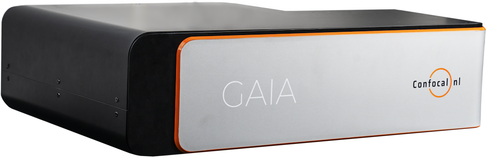
What is GAIA?
GAIA is our Point REscan confocal for live cell imaging in Super resolution. It reaches 170 nm in the raw image and 120 nm after deconvolution, beyond 500 µm deep and over a large field of view, using only microwatts of laser power. Its large pinhole and highly sensitive camera keep phototoxicity very low, so cells can be imaged in Super resolution for longer.
What is the difference between GAIA α and GAIA λ?
GAIA α is the streamlined version, with a fixed pinhole and a single-band filter wheel (quad band optional). GAIA λ is the flagship version, with a switchable pinhole and a motorized single-band filter wheel. Both have the same resolution, speed, field of view and wavelength range.
How fast is GAIA and what field of view does it have?
GAIA images at 3 frames per second at 512 × 512 pixels, and up to 30 frames per second in sprint mode at 256 × 256 pixels. With field number 18 it gives 330 × 330 µm in Super resolution with a 40x objective. It covers visible and near-infrared light, from 400 to 1100 nm.
Which objectives can I use with GAIA?
For Super resolution, use objectives from 30x to 100x; the pinhole covers objectives from 4x to 100x. For the best images, high-NA objectives are a must.
What is the difference between GAIA and the RCM2?
Both are REscan systems with Super resolution of 170 nm raw and 120 nm after deconvolution. GAIA is the newer system: it covers a larger field of view (FN18, 330 × 330 µm with 40x, compared with FN12, 220 × 220 µm, on the RCM2), images faster (3 fps versus 2 fps at 512 × 512 pixels), adds near-infrared up to 1100 nm, and runs in KRONOS.
RCM2
What is the RCM2?
The RCM2 is our second-generation REscan confocal microscope, with digital scanner technology. It turns a widefield microscope into a Super resolution confocal, with 170 nm resolution in the raw image and 120 nm after deconvolution. Its high sensitivity has been demonstrated at only 10 µW excitation power, which minimizes phototoxicity and photobleaching.
What is the difference between the RCM2 and the RCM2.5?
They are the same except for the wavelength range. The RCM2 covers the standard visible range (400 to 700 nm). The RCM2.5 also has an optional visible to near-infrared range (400 to 950 nm).
How fast is the RCM2 and what field of view does it have?
The RCM2 images at 2 frames per second at 512 × 512 pixels, and up to 15 frames per second in sprint mode at 256 × 256 pixels. In Super resolution it has field number 12 (220 × 220 µm with a 40x objective), and field number 18 when not using Super resolution. It comes with a standard quad-band emission filter, with an optional external single-band filter wheel.
What can I do with the RCM2?
The RCM2 offers Super resolution, confocal, widefield and brightfield imaging. It is used for live cell Super resolution and, in combination with silicon objectives, for deep 3D imaging of organoids and zebrafish embryos. Its high signal-to-noise ratio also suits single-molecule detection in low-fluorescence conditions, such as smFISH. The RCM2 runs in Micro-Manager or in your own software with our SDK.
Software
Which software do I use to control the system?
KRONOS is our own acquisition software. It controls the REscan confocal, lasers, camera and microscope in one interface, with GPU rendering, tile stitching and direct export to FIJI. Which other options you have depends on the system:
- NL5+ and AION: KRONOS, ZEISS ZEN with our ZEN Module, Micro-Manager, or our SDK.
- GAIA: KRONOS, Micro-Manager, or our SDK.
- RCM2: Micro-Manager, or our SDK.
What computer do I need for KRONOS?
Minimum: Intel Core i7-13700 processor or higher, 64 GB RAM, NVIDIA GeForce RTX 4060 or Quadro A4000 graphics card, 1 TB NVMe SSD plus a separate backup hard drive, Windows 11, and a display of at least 2560 × 1440 (IPS).
Can I integrate a REscan confocal into my own software?
Yes. The Confocal NL SDK and API is a C++ library (DLL with .lib and .h files) for Windows, with developer guides. It lets you integrate the hardware into third-party or custom software.
Do I need deconvolution?
No, REscan images are ready to use straight away, and Point REscan Super resolution is optical. Deconvolution can improve resolution further, for example from 170 nm to 120 nm on the RCM. For all our systems, deconvolution is done with SVI Huygens as a post-processing step.
Applications
What can I image with a REscan confocal?
Our systems are used for live cell imaging, cell biology, developmental biology, neurobiology, plant biology, Ca²⁺ imaging, C. elegans, organoids, cleared tissue and deep live cell imaging in super-resolution.
Can I image fixed and cleared samples too?
Yes. REscan works for fixed samples as well as living ones. The high sensitivity and the absence of pinhole crosstalk help with large cleared or expanded samples, such as cleared brain tissue and expanded organoids.
Where can I find publications using REscan?
On our Resources page you can find publications, application notes, interviews, webinars and the Quick Knowledge series.
Demos, quotes and support
Can I see a REscan confocal in action or test my own samples?
Yes. Request a demo and our application specialists will arrange a demonstration, where possible with your own samples.
How much does a REscan confocal cost?
The price depends on the system and configuration (microscope, lasers, camera, software). Request a quote and we will send you an offer for your setup.
Do you provide installation and training?
Yes. Our team installs the system and trains your users on site, and our support team stays available afterwards.
How do I get technical support?
Email support@confocalnl.com or use the contact and support form. For sales questions, email sales@confocalnl.com; for anything else, info@confocalnl.com.
Still have a question?
Our application specialists are happy to help you find the right setup for your samples.
Products
Explore our systems
Find the appropriate confocal system for your imaging needs.