
Line REscan in ZEN

Line REscan Confocal now seamlessly integrated into Zeiss ZEN workflow

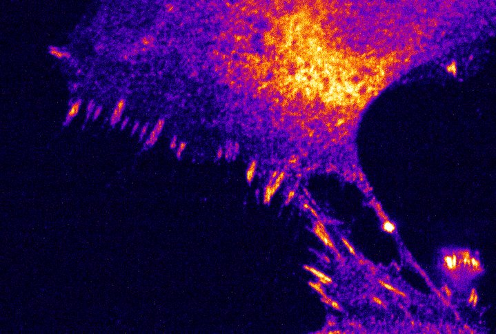
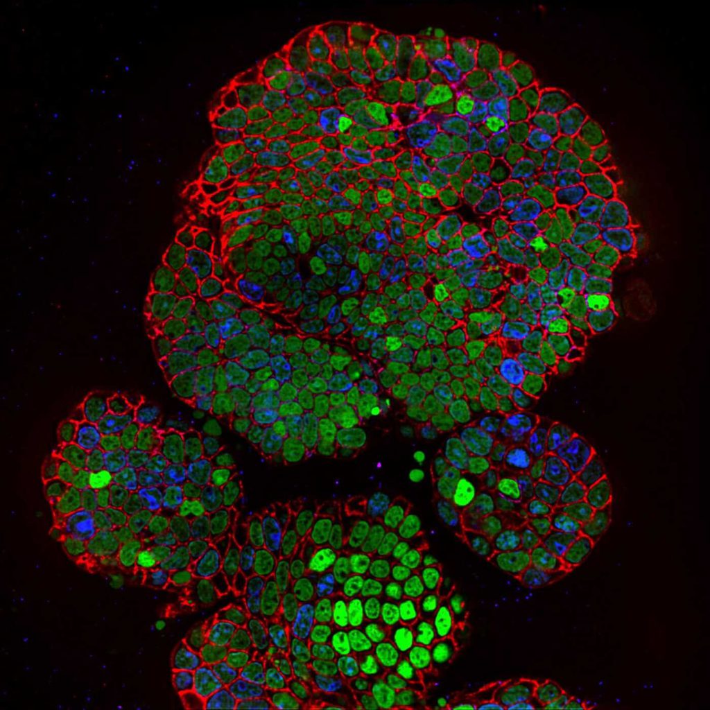
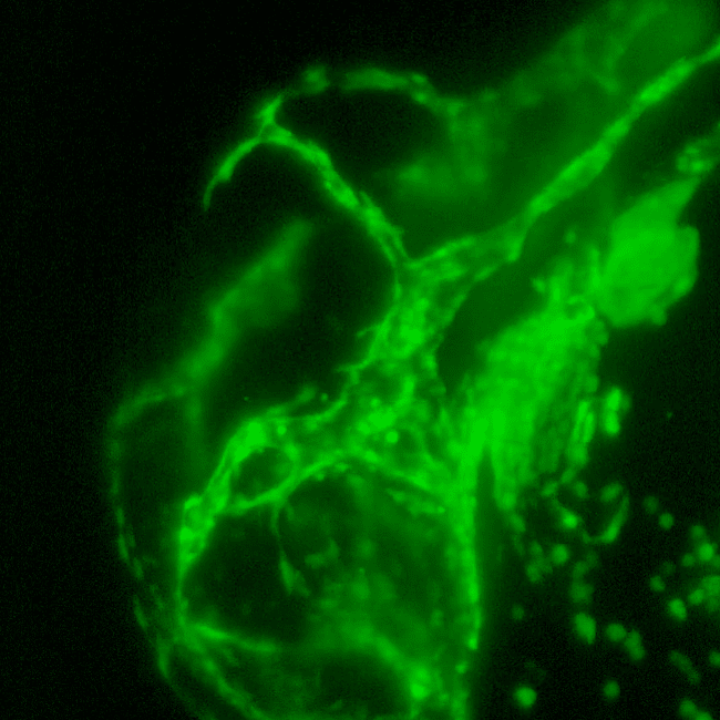
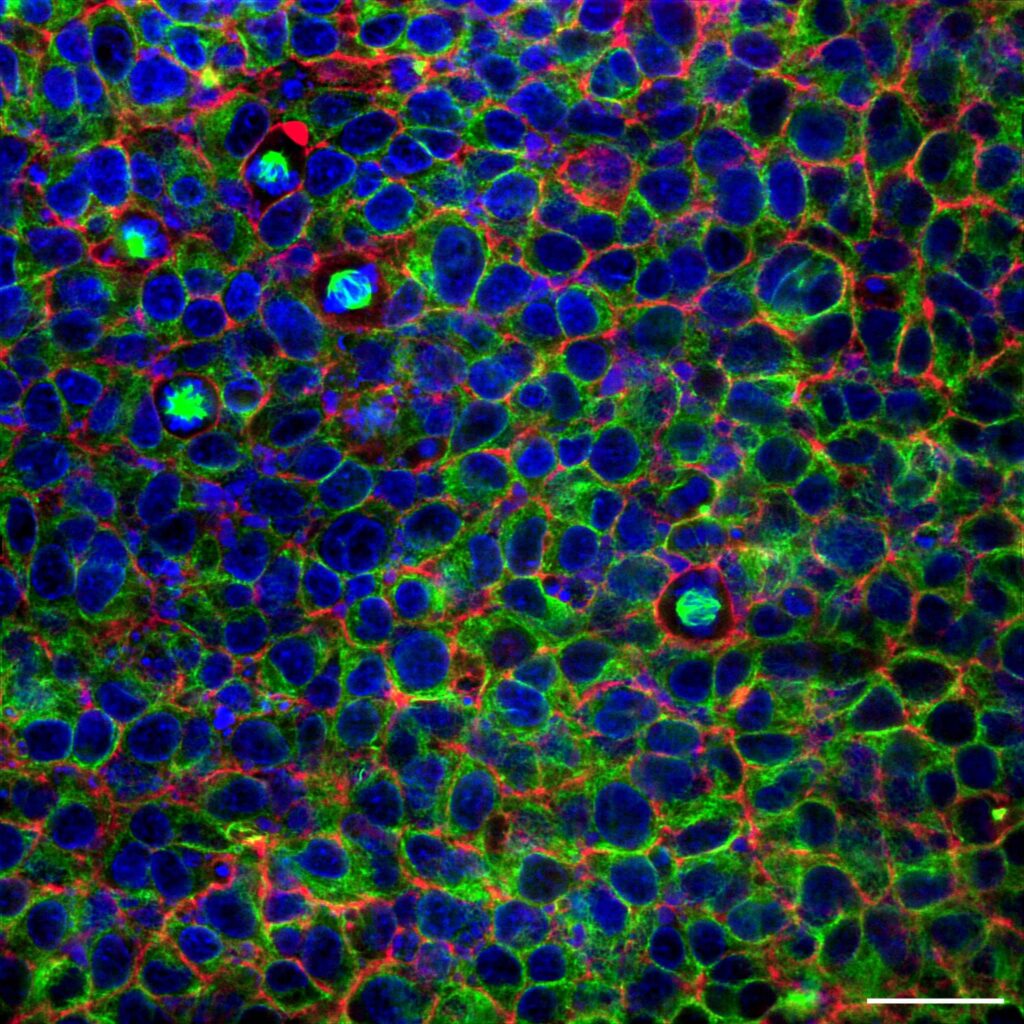
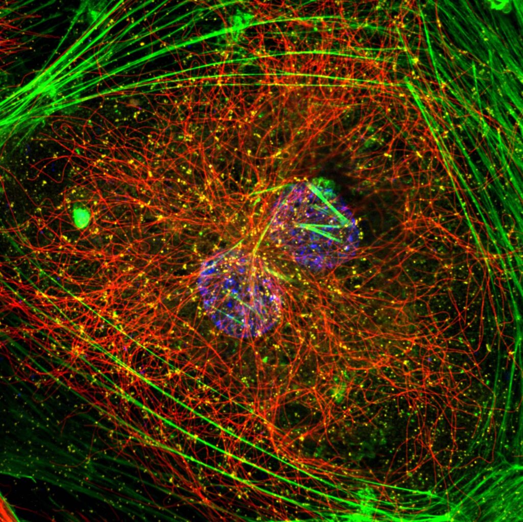
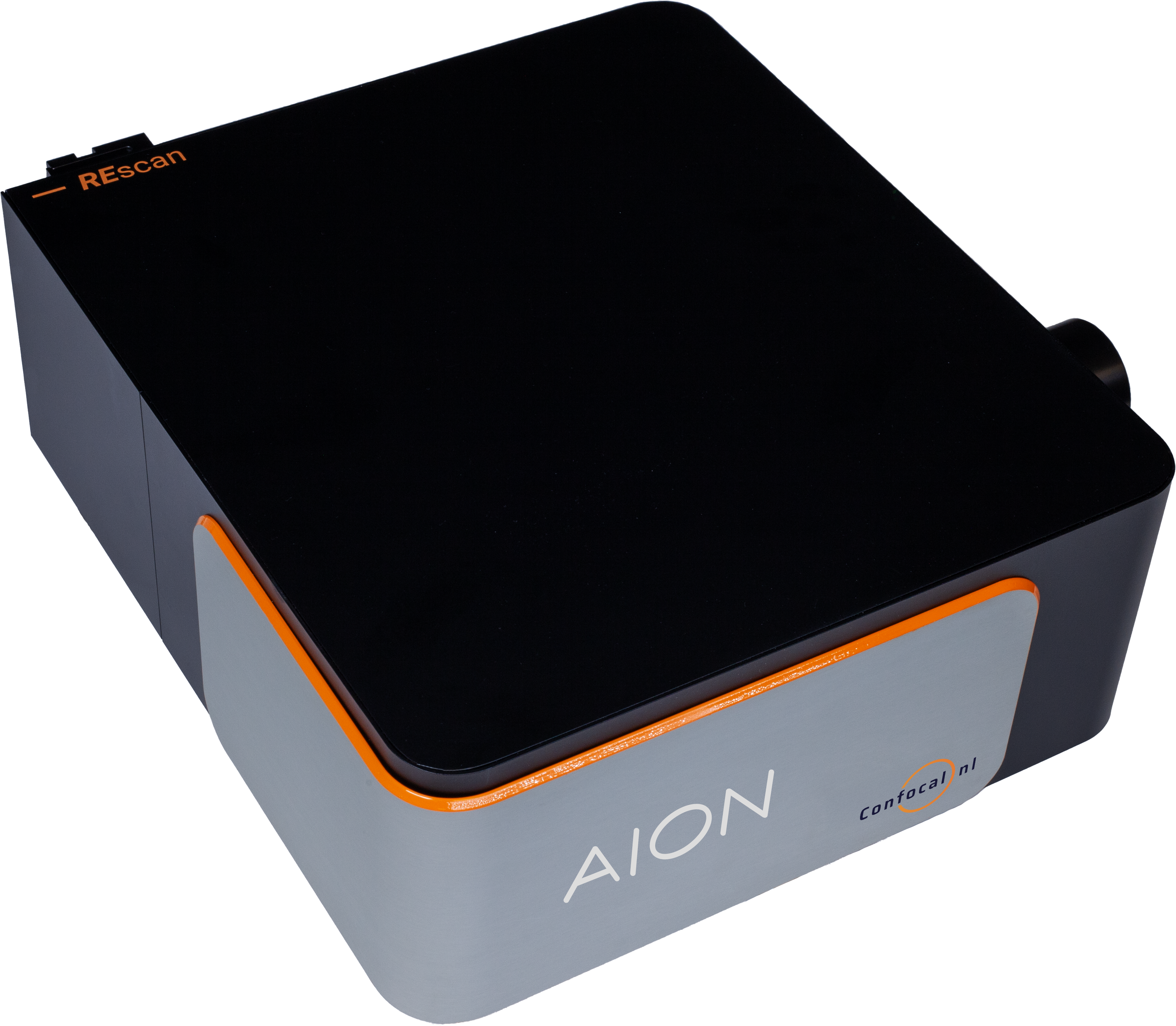
Operate your AION and NL5+ Line REscan confocal microscopes directly within the familiar Zeiss ZEN environment and bring high speed confocal imaging into the workflow researchers already trust. By combining the unique performance of Line REscan technology with the intuitive control, acquisition, and analysis tools of ZEN, users gain a streamlined and powerful imaging platform designed for demanding live cell imaging experiments.

Within a single software ecosystem, researchers can configure imaging parameters, run fast confocal acquisitions, visualize data in real time, and perform advanced analysis without switching between platforms. This tight integration simplifies system operation while preserving the full performance of the Line REscan architecture, delivering exceptional sensitivity, high spatial resolution, and rapid imaging across large fields of view.

Whether capturing fast cellular dynamics, imaging deep into delicate live samples, or running complex multi dimensional experiments, the integration of AION and NL5+ into ZEN removes technical barriers and accelerates discovery. Scientists can work faster, experiment more confidently, and extract deeper insights from their data, all from the interface they already know.

Download flyer

Fast and gentle live cell confocal imaging through ZEN

Full control of your REscan confocal microscope
You can easily control your REscan confocal unit, lasers, cameras, and the microscope body, with seamless switching between confocal, bypass, and widefield modalities.

Control your Line REscan confocal microscope
Easily control your REscan confocal unit, lasers, and cameras, thanks to the seamless integration.

Observe rapid dynamics
Achieve fast acquisition for live samples without switching software environments.

Image deep with close to zero phototoxicity
Reach up to 1 mm deep in your sample with gentle conditions for live samples without switching software environments.

Analyze your data
Use ZEN analysis toolkit either on standalone Line REscan confocal or in combination with Airyscan.

All in ZEN

Acquisition & analysis

Image fast

Rapid cellular dynamics

Go 1 mm deep

Minimal phototoxicity

Reach 95% QE

sCMOS-based detection

NL5+ in ZEN

Deep and fast 3D live cell imaging

  • Capture rapid cellular events at 40 fps at 2048 x 2048 px
  • Observe more with an FN18 field of view on Axio Observer
  • Follow more proteins with a motorized filter wheel with 6 or 7 positions for 25 mm filters
  • Activate a wide range of fluorophores with up to 4 laser lines (400-750 nm)
  • Reach over 1000 fps in fixed line mode (camera limited)
  • Detect with 95% quantum efficiency using a sCMOS camera
  • Utlize motorized widefield/confocal switch
  • Image over days with the lowest phototoxicity in the field

Learn more about the NL5+

AION in ZEN

The new standard in confocal microscopy

  • Everything the NL5+ can do… only more so
  • The only live cell imaging confocal with a switchable pinhole
  • Obtain perfect confocality with any objective
  • Automatic slit-shaped pinhole selection providing ~ 1,5 Airy Units depending on the wavelength and the objective
  • Capture the fastest dynamic events in the cell at >130 fps at 2304 x 2304 px
  • See more than ever on Axio Observer using FN22 field of view
  • Activate the widest range of fluorophores with up to 8 lines 400-1100 nm (VIS & NIR)
  • Image even the most sensitive samples over days using the widest open pinhole without affecting the resolution

Learn more about the AION

Applications

Cell biology
Visualize cells and their internal structures in high detail, from organelles to individual proteins, using confocal and Super resolution microscopy to support breakthroughs in cell biology and drug discovery.

Developmental biology
Follow cell differentiation and organ formation as they unfold, while investigating the signalling pathways that guide development.

Live cell imaging
Examine living cells in real time with optical sectioning and time-lapse microscopy, capturing dynamic processes such as division, migration, trafficking, and signalling while maintaining cell viability.

Deep Live cell imaging in Super Resolution
Go beyond the diffraction limit with Super resolution imaging to uncover nanoscale structures, protein interactions, and dynamic cellular processes that deepen insights into health and disease.

Neurobiology
Map synaptic connections with exceptional precision using confocal and super-resolution microscopy, revealing nanoscale organization and protein localization to advance understanding of brain function and neurological disorders.

Plant Biology
Explore intricate three dimensional plant structures with confocal microscopy, from root systems to leaf tissues, while monitoring processes like photosynthesis, nutrient flow, and cell division.

Ca²⁺ imaging
Track real-time fluctuations in intracellular calcium levels to uncover essential signalling mechanisms, particularly in neuroscience and cardiac research.

C. elegans
Investigate fundamental biological processes in C. elegans by imaging its transparent body in real time, enabling studies of development, behavior, neural circuitry, gene expression, and synaptic structure.

Organoids
Study cell interactions within physiologically relevant environments by imaging organoids, supporting research into development, disease modeling, drug testing, and personalized medicine.

Cleared tissue
Examine intact biological systems in three dimensions using tissue clearing techniques, preserving structural integrity while allowing deep imaging of complex networks such as neurons and vasculature.

Resources

Related Resources

Download the product flyer

Receive more information on this product or request a quote.

Download now